

Ultra-High-Resolution Photon-Counting Detector CT Benefits Visualization of Abdominal Arteries: A Comparison to Standard-Reconstruction
- PMID: 39455541
- PMCID: PMC12092866
- DOI: 10.1007/s10278-024-01232-5
Ultra-High-Resolution Photon-Counting Detector CT Benefits Visualization of Abdominal Arteries: A Comparison to Standard-Reconstruction
Abstract
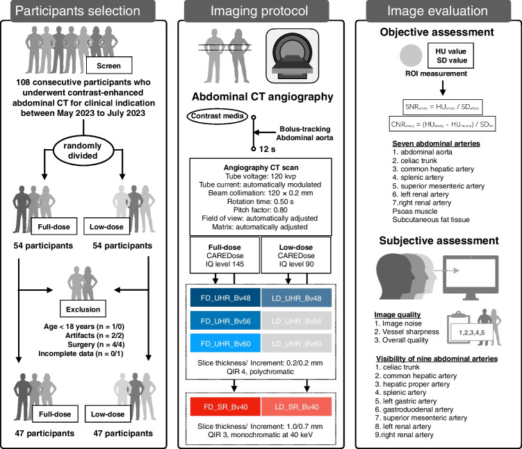
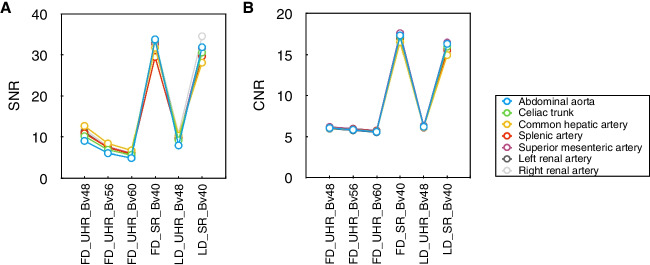
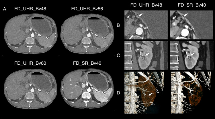
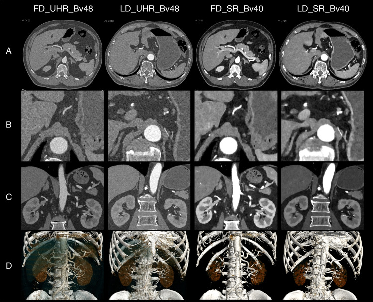
This study aimed to investigate the potential benefit of ultra-high-resolution (UHR) photon-counting detector CT (PCD-CT) angiography in visualization of abdominal arteries in comparison to standard-reconstruction (SR) images of virtual monoenergetic images (VMI) at low kiloelectron volt (keV). We prospectively included 47 and 47 participants to undergo contrast-enhanced abdominal CT scans within UHR mode on a PCD-CT system using full-dose (FD) and low-dose (LD) protocols, respectively. The data were reconstructed into six series of images: FD_UHR_Br48, FD_UHR_Bv56, FD_UHR_Bv60, FD_SR_Bv40, LD_UHR_Bv48, and LD_SR_Bv40. The UHR reconstructions were performed with three kernels (Bv48, Bv56, and Bv60) within 0.2 mm. The SR were virtual monoenergetic imaging reconstruction with Bv40 kernel at 40-keV within 1 mm. Each series of axial images were reconstructed into coronal and volume-rendered images. The signal-to-noise ratio (SNR) and contrast-to-noise ratio (CNR) of seven arteries were measured. Three radiologists assessed the image quality, and visibility of nine arteries on all the images. SNR and CNR values of SR images were significantly higher than those of UHR images (P < 0.001). The SR images have higher ratings in image noise (P < 0.001), but the FD_UHR_Bv56 and FD_UHR_Bv60 images has higher rating in vessel sharpness (P < 0.001). The overall quality was not significantly different among FD_VMI_40keV, LD_VMI_40keV, FD_UHR_Bv48, and LD_UHR_Bv48 images (P > 0.05) but higher than those of FD_UHR_Bv56 and FD_UHR_Bv60 images (P < 0.001). There is no significant difference of nine abdominal arteries among six series of images of axial, coronal and volume-rendered images (P > 0.05). To conclude, 1-mm SR image of VMI at 40-keV is superior to 0.2-mm UHR regardless of which kernel is used to visualize abdominal arteries, while 0.2-mm UHR image using a relatively smooth kernel may allow similar image quality and artery visibility when thinner slice image is warranted.
Keywords: Computed tomography angiography; Contrast media; Image enhancement; Image reconstruction; Radiation dosage.
© 2024. The Author(s).
Conflict of interest statement
Declarations. Ethics Approval: This study was performed in line with the principles of the Declaration of Helsinki. Institutional Review Board approval was obtained from Ruijin Hospital, Shanghai Jiao Tong University School of Medicine (No. 2015–76). Consent to Participate: Written informed consent from all participants were received. Consent for Publication: The authors affirm that human research participants provided informed consent for publication of the images of Fig 5 and Fig6 andthose in the Supplementary Material. Competing Interests: Ms. Zhihan Xu is an employee of Siemens Healthineers, which is the manufacturer of the CT system used in this study. However, she neither had access nor control on the data acquisition and analysis. Dr. Jingyu Zhong acknowledges his position as a member of the Scientific Editorial Board of European Radiology and BMC Medical Imaging. All other authors of this manuscript declare no relationships with any companies, whose products or services may be related to the subject matter of the article. Overlapping Cohort: The abstract of this study entitled “Does ultra-high-resolution photon-counting detector CT benefit visualization of abdominal arteries? A comparison to standard-reconstruction” (C-22800) has been accepted as a digital poster, EPOS Radiologist (scientific), on European Congress of Radiology 2024 ( https://doi.org/ https://doi.org/10.26044/ecr2024/C-22800 ). The presenting author of this abstract is Dr. Jingyu Zhong.
Figures


References
-
- Fleischmann D, Afifi RO, Casanegra AI et al; American Heart Association Council on Cardiovascular Radiology and Intervention; Council on Arteriosclerosis, Thrombosis and Vascular Biology; Council on Clinical Cardiology; Council on Cardiovascular Surgery and Anesthesia (2022) Imaging and surveillance of chronic aortic dissection: a scientific statement from the American Heart Association. Circ Cardiovasc Imaging 15(3):e000075. 10.1161/HCI.0000000000000075 - PubMed
Publication types
MeSH terms
Substances
Grants and funding
- 82302183/National Natural Science Foundation of China
- 82271934/National Natural Science Foundation of China
- 22YF1442400/Yangfan Project of Science and Technology Commission of Shanghai Municipality
- 2023QN01/Research Found of Health Commission of Changing District, Shanghai Municipality
- 2024JZWC-YBA07/Laboratory Open Fund of Key Technology and Materials in Minimally Invasive Spine Surgery
- TRKYRC-XX202204/Research Fund of Tongren Hospital, Shanghai Jiao Tong University School of Medicine
- TRYJ2021JC06/Research Fund of Tongren Hospital, Shanghai Jiao Tong University School of Medicine
- TRYXJH18/Research Fund of Tongren Hospital, Shanghai Jiao Tong University School of Medicine
- TRYXJH28/Research Fund of Tongren Hospital, Shanghai Jiao Tong University School of Medicine
LinkOut - more resources
Full Text Sources
Medical
Research Materials