. 2024 Oct 26;15(1):9244.
doi: 10.1038/s41467-024-53639-x.

Regulation of hepatic inclusions and fibrinogen biogenesis by SEL1L-HRD1 ERAD

Affiliations

Regulation of hepatic inclusions and fibrinogen biogenesis by SEL1L-HRD1 ERAD

Zhenfeng Song et al. Nat Commun. .

Abstract

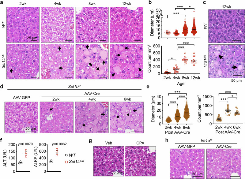
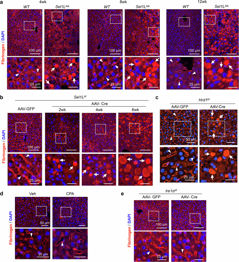
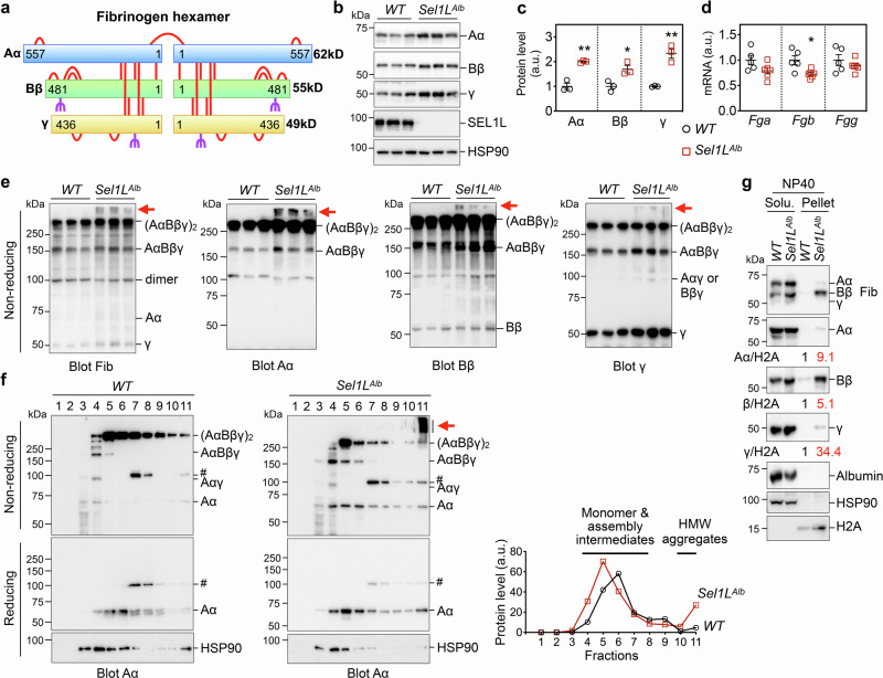
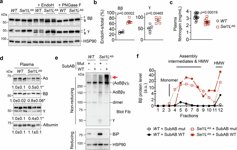
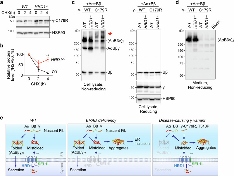
Impaired secretion of an essential blood coagulation factor fibrinogen leads to hepatic fibrinogen storage disease (HFSD), characterized by the presence of fibrinogen-positive inclusion bodies and hypofibrinogenemia. However, the molecular mechanisms underlying the biogenesis of fibrinogen in the endoplasmic reticulum (ER) remain unexplored. Here we uncover a key role of SEL1L-HRD1 complex of ER-associated degradation (ERAD) in the formation of aberrant inclusion bodies, and the biogenesis of nascent fibrinogen protein complex in hepatocytes. Acute or chronic deficiency of SEL1L-HRD1 ERAD in the hepatocytes leads to the formation of hepatocellular inclusion bodies. Proteomics studies followed by biochemical assays reveal fibrinogen as a major component of the inclusion bodies. Mechanistically, we show that the degradation of misfolded endogenous fibrinogen Aα, Bβ, and γ chains by SEL1L-HRD1 ERAD is indispensable for the formation of a functional fibrinogen complex in the ER. Providing clinical relevance of these findings, SEL1L-HRD1 ERAD indeed degrades and thereby attenuates the pathogenicity of two disease-causing fibrinogen γ mutants. Together, this study demonstrates an essential role of SEL1L-HRD1 ERAD in fibrinogen biogenesis and provides insight into the pathogenesis of protein-misfolding diseases.


Conflict of interest statement

The authors declared no competing interests.

Figures

Fig. 1
Fig. 1. Formation of inclusion bodies in Sel1L-deficient livers.
a–b Representative images of H&E stained liver sections of male WT (Sel1Lf/f) and Sel1LAlb (Sel1Lf/f;Albumin-Cre) littermates at different ages. The size and number of inclusion bodies found in Sel1LAlb livers are quantitated in b. N = 3 per cohort. c Representative images of H&E stained liver sections of 12-week-old WT and Hrd1Alb littermates. N = 3. d–e Sel1Lf/f mice were injected i.v. with AAV8 expressing hepatocyte-specific TBG promoter-driven Cre or GFP. Representative images of H&E-stained liver sections of mice at different time points after the injection are shown d. The size and number of inclusion bodies are quantitated in e. N = 4 for GFP cohort, n = 3 for 2 wk and 4 wk post-AAV-Cre, n = 5 for 6 wk post-AAV-Cre. In (a), (c) and (d), arrows point to inclusion bodies. f Serum from 6-week-old WT and Sel1LAlb littermates were analyzed for alanine aminotransferase (ALT) and alkaline phosphatase (ALKP). N = 3. g–h Representative images of H&E stained liver sections of (g) WT mice 4 weeks after i.p. injections of vehicle (Veh) or cyclopiazonic acid (CPA) at 5 mg/kg body weight every other day, n = 3; and (h) Ire1αf/f mice 6 weeks after an i.v. injection of AAV8 expressing TBG-driven Cre or GFP, n = 5. For inclusion body quantitation, 12 to 16 images taken from indicated number of mice per cohort were analyzed. Values, mean ± SEM. *, p < 0.05; ***, p < 0.001 by one-way ANOVA with Tukey multiple comparison test in (b) and (e), and by two-tailed Student’s t test in f. Source data are provided as a Source Data file.
Fig. 2
Fig. 2. Inclusion bodies are encased in ER membrane with the identification of fibrinogen chains as a possible major component.
a–d TEM images of liver tissues from 6-week-old WT and Sel1LAlb littermates with insets of higher magnification shown (n = 2 mice each genotype). White arrowheads, normal ER; red arrows, inclusion bodies bounded by a single membrane; white arrows, dilated ER; green arrow, a vesicle in the process of fusing to a large inclusion body; N, nucleus; m, mitochondria; IB, inclusion bodies; LD, lipid droplets. Glycogen granules are noted in both WT and Sel1LAlb livers. e Proteomics analysis of purified microsomes from WT and Sel1LAlb livers, showing the fibrinogen chains significantly over-represented in the Sel1LAlb samples. Values represent scaled abundances of proteins from tandem mass tag (TMT) labeling-based mass spectrometry. P values were caculated by two-tailed Student’s t test without adjustments for multiple comparisons.
Fig. 3
Fig. 3. Fibrinogen is largely trapped in the inclusion bodies of SEL1L-HRD1 ERAD-deficient hepatocytes.
Fibrinogen staining using a rabbit anti-serum recognizing all chains of fibrinogen in liver sections from: (a) WT and Sel1LAlb littermates at different ages; (b) Sel1Lf/f mice at different time points after an i.v. injection of AAV8 expressing TBG-driven Cre or GFP; (c) Hrd1f/f mice 4 weeks after an i.v. injection of AAV8-TBG-Cre or GFP; (d) WT mice 4 weeks after i.p. injections of vehicle (Veh) or cyclopiazonic acid (CPA) at 5 mg/kg body weight every other day; (e) Ire1αf/f mice 6 weeks after an i.v. injection of AAV8-TBG-Cre or GFP. Insets of higher magnification shown below. Arrowheads point to apical and sinusoidal localization of fibrinogen; arrows point to inclusion bodies containing fibrinogen. Representative data from n = 3 mice per cohort.
Fig. 4
Fig. 4. Fibrinogen is in a misfolded or folding intermediate state in Sel1L-deficient hepatocytes.
a Primary hepatocytes were isolated from WT and Sel1LAlb littermates and stained with the indicated antibodies. Note the co-localizations of fibrinogen and an ER marker KDEL (recognizing BiP and GRP94) in the inclusion bodies of Sel1LAlb hepatocytes (white arrows). White dotted lines outline individual hepatocytes. Representative data from two independent repeats. b, c TEM couple to Immunogold labeling of BiP in WT and Sel1LAlb livers. White arrows mark the localization of BiP in normal ER in WT hepatocytes. Representitave image of eight hepatocytes from one mouse per genotype shown. Red arrows mark BiP in inclusion bodies in Sel1LAlb hepatocytes. White dotted lines mark the boundary of inclusion bodies. N, nucleus; m, mitochondria.
Fig. 5
Fig. 5. Fibrinogen chains accumulate and form insoluble aggregates in Sel1L-deficient livers.
a The overall structure of fibrinogen showing a hexamer of Aα, Bβ and γ chains. The 29 highly conserved disulfide bonds are shown as red bars, and the glycosylation sites on Bβ and γ are marked in purple. Aα is not glycosylated. Image created in BioRender. Tushi, N. (2024) BioRender.com/l49l811. (b–e) Liver samples from 6-week-old WT and Sel1LAlb littermates were analyzed for: (b) reducing Western blot analysis with quantitation normalized to HSP90 shown in (c); (d) qPCR analysis; (e) non-reducing SDS-PAGE and Western blot analysis using the same protein lysates shown in (b). In (b) and (e), fibrinogen proteins were analyzed by antibodies specific for each chain or all fibrinogen chains (Fib). Red arrows point to high molecular weight aggregates of fibrinogen. N = 3 for Western blot analyses, n = 5 for WT and n = 6 for Sel1LAlb for qPCR analyses. Values, mean ± SEM. *, p < 0.05; **, p < 0.01 by two-tailed Student’s t test. f Sucrose gradient fractionation (fractions 1 to 11 from top to bottom) of liver samples from WT and Sel1LAlb littermates analyzed by nonreducing or reducing SDS-PAGE using an Aα chain-specific antibody. Red arrow points to fibrinogen aggregates. #, a non-specific band. Quantitation of reduced Aα is shown on the right with fractions with Aα monomers, assembly intermediates and high molecular weight (HMW) aggregates labeled. HSP90, a loading control. g Liver samples from 6-week-old WT and Sel1LAlb littermates were prepared in lysis buffer containing 0.5% Nonidet P-40 (NP40) and analyzed for protein distribution in the NP40 soluble and NP40 insoluble (pellet) fractions by Western blot. The distribution of HSP90 and H2A marks the soluble and insoluble fractions, respectively. Quantitation of fibrinogen chains normalized to H2A in NP40 insoluble fraction is shown below the blots. (f–g), representative data from two independent repeats. Source data are provided as a Source Data file.
Fig. 6
Fig. 6. Fibrinogen is retained in the ER of Sel1L-deficient hepatocytes, with BiP attenuating its further aggregation.
a–b Western blot analysis of fibrinogen Bβ and γ in liver lysates treated with EndoH or PNGase F. R and s, EndoH-resistant and sensitive, respectively. The percentages of EndoH-sensitive Bβ and γ chains are quantitated in b. N = 5 per cohort. c–d ELISA (c) and Western blot (d) analyses of inferior vena cava (IVC) plasma from 6-week-old WT and Sel1LAlb littermates. Quantitation of Western blot is shown below the blots. WT, n = 15; Sel1LAlb, n = 10 for ELISA; n = 3 for Western blot. Values, mean ± SEM. *, p < 0.05 or as labeled by two-tailed Student’s t test. (e-f) Primary hepatocytes isolated from WT and Sel1LAlb littermates were treated with 1 µg/mL WT SubAB or mutant SubAA272B (mut) for 12 hours and analyzed by: (e) non-reducing and reducing SDS-PAGE followed by Western blot analyses, with red arrow pointing to fibrinogen aggregates; and (f) sucrose gradient fractionation (fractions 1 to 12 from top to bottom) showing the quantitation of reduced Bβ chain, with monomers, assembly intermediates and HMW aggregates labeled. Western blot images for (f) are shown in Fig. S7c. Representative data from two independent repeats. Source data are provided as a Source Data file.
Fig. 7
Fig. 7. Three fibrinogen chains are bona fide endogenous ERAD substrates.
a Western blot analysis of immunoprecipitates of HA-agarose in HEK293T cells transfected with indicated plasmids, showing that HRD1 is sufficient for the ubiquitination of fibrinogen chains. HRD1-C2A, an E3 ligase dead mutant. Cells were treated with a proteasome inhibitor MG132 for the last 3 hr prior to immunoprecipitation. Representative data from two independent repeats. b–c Western blot analysis of endogenous fibrinogen Aα, Bβ and γ decay after a translation inhibitor cycloheximide (CHX) treatment in (b) primary hepatocytes isolated from WT and Sel1LAlb littermates, and (c) WT, SEL1L−/− and HRD1−/− Huh7 hepatocytes. The degradation is quantitated relative to the percentage of control. In (b), n = 4 for Aα and Bβ; n = 3 for γ each genotype. In c, n = 5 for WT and HRD1−/−; n = 3 for SEL1L−/−. N indicates independent samples. Values, mean ± SEM. *, p < 0.05; **, p < 0.01; ***, p < 0.001 comparing knockout to WT cells by two-way ANOVA with Sidak multiple comparison test. Source data are provided as a Source Data file.
Fig. 8
Fig. 8. Assembly of fibrinogen complex prevents SEL1L-HRD1 ERAD of individual chains.
8-week-old WT and Sel1LAlb littermates were injected intravenously with lipid nanoparticle (LNP)-encapsulated small interfering RNA against fibrinogen Aα chain (siFga) or luciferase (siLuc) for control. 11 days after injection, mice were examined for: (a) reducing Western blot analysis of liver samples with quantitation normalized to HSP90 shown on the right; (b) non-reducing SDS-PAGE and Western blot analysis using the same liver lysates shown in a; (c) Western blot analysis of liver lysates treated with EndoH, with the percentages of EndoH-sensitive Bβ and γ chains shown on the right; and (d) H&E and fibrinogen staining of liver sections, with the size of inclusion bodies in H&E-stained Sel1LAlb samples quantitated on the right. N = 3. In (d), 12 to 16 images taken from 3 mice per cohort were analyzed. Values, mean ± SEM. *, p < 0.05; ***, p < 0.001 by two-way ANOVA with Tukey multiple comparison test in (a) and (c), and by student’s t test in d. Source data are provided as a Source Data file.
Fig. 9
Fig. 9. SEL1L-HRD1 ERAD degrades a disease-causing fibrinogen γ variant and mitigates its pathogenicity.
a–b Western blot analysis of fibrinogen γ-C179R decay after a translation inhibitor CHX treatment in WT and HRD1−/− HEK293T cells transfected with a plasmid expressing γ-C179R. The degradation is quantitated relative to the percentage of control. N = 3, indicating independent samples. Values, mean ± SEM. **, p < 0.01 by two-way ANOVA with Sidak multiple comparison test. c–d WT and HRD1−/− HEK293T cells were transfected with Aα-WT, Bβ-WT, and either γ-WT or γ-C179R. Cell lysates (c) and cell culture medium (d) were analyzed by non-reducing or reducing SDS-PAGE as indicated. Red arrow points to fibrinogen Bβ aggregates. Culture medium from non-transfected cells was included as a blank control in d. Representative data from two independent repeats. e Model showing a critical role of SEL1L-HRD1 ERAD in the biogenesis of mature fibrinogen protein complex in the ER. SEL1L-HRD1 ERAD in hepatocytes mediates the degradation of endogenous fibrinogen chains, thereby ensuring their proper assembly and subsequent secretion (left). In the absence of ERAD, fibrinogen chains accumulate and aggregate in ER-derived hepatic inclusion bodies, interfering the folding and secretion of nascent fibrinogen protein (middle). SEL1L-HRD1 ERAD also degrades a disease-causing fibrinogen γ mutant, thereby attenuating its pathogenicity (right). Image created in BioRender. Tushi, N. (2024) BioRender.com/k40b037. Source data are provided as a Source Data file.

References

    1. Pieters, M. & Wolberg, A. S. Fibrinogen and fibrin: An illustrated review. Res Pr. Thromb. Haemost.3, 161–172 (2019). - PMC - PubMed
    1. Luyendyk, J. P., Schoenecker, J. G. & Flick, M. J. The multifaceted role of fibrinogen in tissue injury and inflammation. Blood133, 511–520 (2019). - PMC - PubMed
    1. Liu, X. & Shi, B. Progress in research on the role of fibrinogen in lung cancer. Open Life Sci.15, 326–330 (2020). - PMC - PubMed
    1. Casini A., Neerman-Arbez M., & de Moerloose P. Heterogeneity of congenital afibrinogenemia, from epidemiology to clinical consequences and management. Blood Rev. 100793 (2020). - PubMed
    1. Tamura, T., Arai, S., Nagaya, H., Mizuguchi, J. & Wada, I. Stepwise assembly of fibrinogen is assisted by the endoplasmic reticulum lectin-chaperone system in HepG2 cells. PLoS One8, e74580 (2013). - PMC - PubMed

Publication types

MeSH terms

LinkOut - more resources