. 2024 Oct 25;15(1):9223.
doi: 10.1038/s41467-024-53587-6.

Mefloquine-induced conformational shift in Cx36 N-terminal helix leading to channel closure mediated by lipid bilayer

Affiliations

Mefloquine-induced conformational shift in Cx36 N-terminal helix leading to channel closure mediated by lipid bilayer

Hwa-Jin Cho et al. Nat Commun. .

Abstract

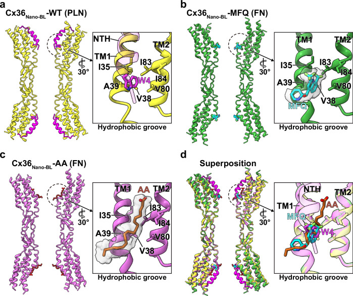
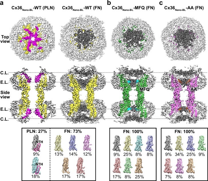
Connexin 36 (Cx36) forms interneuronal gap junctions, establishing electrical synapses for rapid synaptic transmission. In disease conditions, inhibiting Cx36 gap junction channels (GJCs) is beneficial, as it prevents abnormal synchronous neuronal firing and apoptotic signal propagation, mitigating seizures and progressive cell death. Here, we present cryo-electron microscopy structures of human Cx36 GJC in complex with known channel inhibitors, such as mefloquine, arachidonic acid, and 1-hexanol. Notably, these inhibitors competitively bind to the binding pocket of the N-terminal helices (NTH), inducing a conformational shift from the pore-lining NTH (PLN) state to the flexible NTH (FN) state. This leads to the obstruction of the channel pore by flat double-layer densities of lipids. These studies elucidate the molecular mechanisms of how Cx36 GJC can be modulated by inhibitors, providing valuable insights into potential therapeutic applications.


Conflict of interest statement

The authors declare no competing interests.

Figures

Fig. 1
Fig. 1. Cryo-EM structures of human Cx36 in complex with mefloquine and arachidonic acid.
a A schematic diagram summarizing the seven cryo-EM structures of human Cx36 in this study, including the nomenclature used. b, c Cryo-EM maps representing surface hydrophobicity of Cx36Nano-BL-MFQ and Cx36Nano-BL-AA in cross-sectioned side view. The upper and bottom boxes present close-ups of the hydrophobic groove and a top view, respectively. The water-accessible pore diameter is indicated in the bottom boxes. The surface hydrophobicity (Log P) of atomic models is computed and visualized using UCSF ChimeraX, where hydrophobic and hydrophilic surfaces are color-coded in a gradient of dark gray and blue. The channel pore-bound MFQ and AA are depicted in cyan and brown sticks, respectively. d A comparison of the water-accessible pore diameter of Cx36 GJC structures is shown. Cx36Nano-BL-WT in PLN and FN states are colored in black and yellow dashed lines, while Cx36Nano-BL-MFQ and Cx36Nano-BL-AA are colored in green and purple solid lines, respectively. Source data are provided as a Source Data file.
Fig. 2
Fig. 2. Common hydrophobic binding site of MFQ and AA in Cx36 GJC.
ad The ribbon representation of Cx36Nano-BL-WT in PLN (a yellow ribbon), Cx36Nano-BL-MFQ (b green ribbon), Cx36Nano-BL-AA (c pink ribbon), and their superimposition (d). The close-up of the H.G. is presented in boxes. The H.G. is composed of hydrophobic residues from TM1 (Ile35, Val38, and Ala39) and TM2 (Val80, Ile83, and Ile84). The NTH and Trp4 of Cx36Nano-BL-WT in PLN are colored in magenta. H.G.-bound MFQ and AA, along with their coulomb density maps, are shown in cyan, brown, and gray, respectively. H.G. hydrophobic groove, NTH N-terminal helix, PLN pore-lining NTH, FN flexible NTH, MFQ mefloquine, AA arachidonic acid.
Fig. 3
Fig. 3. Structural characterization of the pore-occluded states of Cx36Nano-BL-MFQ and Cx36Nano-BL-AA in lipid nanodiscs.
ac Top and cross-sectioned side views of the cryo-EM reconstruction map with C1 symmetry imposition. The densities of Cx36Nano-BL-WT in PLN and FN states (a) are displayed in yellow, while Cx36Nano-BL-MFQ (b) and Cx36Nano-BL-AA (c) are displayed in green and pink, respectively. The NTHs and double-layered pore-occluding lipids at the C.L. and E.L. are highlighted in magenta and dark gray, respectively. The hydrophobic groove (H.G.)-bound MFQ and AA, as well as lipid nanodiscs, are shown in cyan, brown, and white, respectively. The summary of the protomer-focused 3D classification of Cx36Nano-BL-WT, Cx36Nano-BL-MFQ, and Cx36Nano-BL-AA is viewed in the bottom boxes. NTH N-terminal helix, PLN pore-lining NTH, FN flexible NTH, MFQ mefloquine, AA arachidonic acid, C.L. cytoplasmic layer, E.L. extracellular layer.
Fig. 4
Fig. 4. The unfavorable binding of MFQ to the H.G. in the FN state of NTH of Cx36 in lipid nanodiscs.
a Structural comparison around the hydrophobic pocket of Cx36Nano-BL-WT (yellow) with the PLN state and the previously determined structure of Cx36-WT in soybean lipids (Cx36Nano-SL-WT, gray). The NTH is colored in magenta. The circled N, 1, and 2 denote NTH, TM1, and TM2, respectively. b, c Top and cross-sectioned side views of the cryo-EM reconstruction map with C1 symmetry imposition. The GJC densities of Cx36Nano-BL-ΔA14-MFQ (b) and Cx36Nano-BL-ΔA14 (c) are displayed in dark red and purple, respectively. The double-layered pore-occluding lipids at the C.L. and E.L. and lipid nanodiscs are colored dark gray and white, respectively. The close-ups of H.G. of Cx36Nano-BL-MFQ and Cx36Nano-BL-ΔA14-MFQ are presented in boxes. The ribbon representation of Cx36Nano-BL-MFQ and Cx36Nano-BL-ΔA14-MFQ GJC, and their additional densities at H.G. are colored in green, salmon, cyan, and white, respectively. The additional densities are contoured at 4 σ.
Fig. 5
Fig. 5. Schematic model for channel inhibition of Cx36 GJC by chemical inhibitors.
Schematic representation of the open and pore-occluded states of Cx36 GJC. The NTH, TM1 in the PLN and FN states, TM2-4 and ECL1-2, lipids in the channel pore, lipid bilayer, and cation are colored magenta, yellow, green, gray, dark gray, light gray, and sky blue, respectively. Trp4 of PLN and MFQ are highlighted in green and blue, respectively. MFQ competitively binds to the H.G., where the NTH binds through Trp4. This induces a conformational change from PLN to FN state, shifting the conformational dynamic equilibrium of Cx36 GJC toward the FN state.

Similar articles

References

    1. Laird, D. W. & Lampe, P. D. Therapeutic strategies targeting connexins. Nat. Rev. Drug Discov.17, 905–921 (2018). - PMC - PubMed
    1. Brücher, B. L. & Jamall, I. S. Cell-cell communication in the tumor microenvironment, carcinogenesis, and anticancer treatment. Cell. Physiol. Biochem.34, 213–243 (2014). - PubMed
    1. Totland, M. Z. et al. Regulation of gap junction intercellular communication by connexin ubiquitination: physiological and pathophysiological implications. Cell. Mol. Life Sci.77, 573–591 (2020). - PMC - PubMed
    1. Bargiello, T. A. et al. Gating of Connexin Channels by transjunctional-voltage: conformations and models of open and closed states. Biochim. Biophys. Acta1860, 22–39 (2018). - PMC - PubMed
    1. Goodenough, D. A. & Paul, D. L. Beyond the gap: functions of unpaired connexon channels. Nat. Rev. Mol. Cell Biol.4, 285–295 (2003). - PubMed

Publication types

LinkOut - more resources