. 2024 Oct 25;9(1):48.
doi: 10.1038/s41525-024-00429-5.

Biallelic GGGCC repeat expansion leading to NAXE-related mitochondrial encephalopathy

Affiliations

Biallelic GGGCC repeat expansion leading to NAXE-related mitochondrial encephalopathy

Kokoro Ozaki et al. NPJ Genom Med. .

Abstract

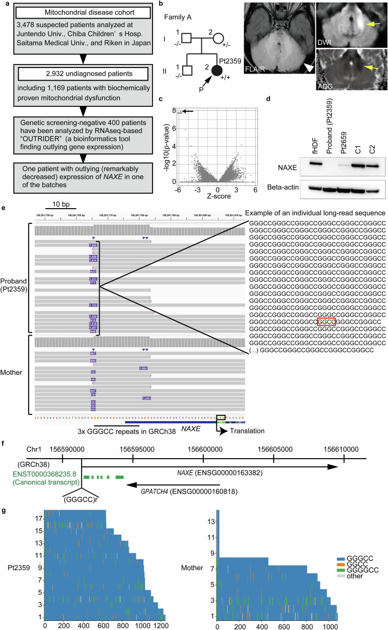
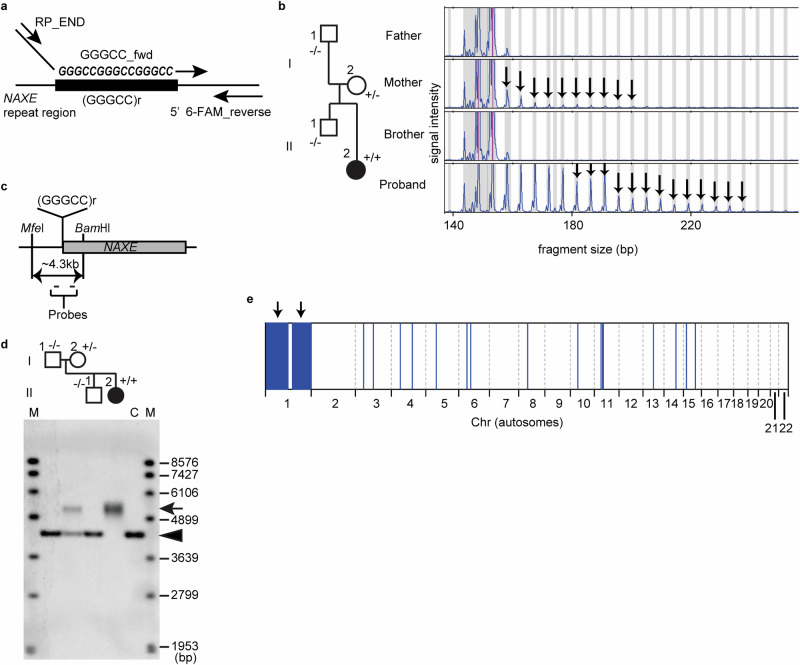
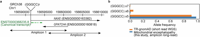
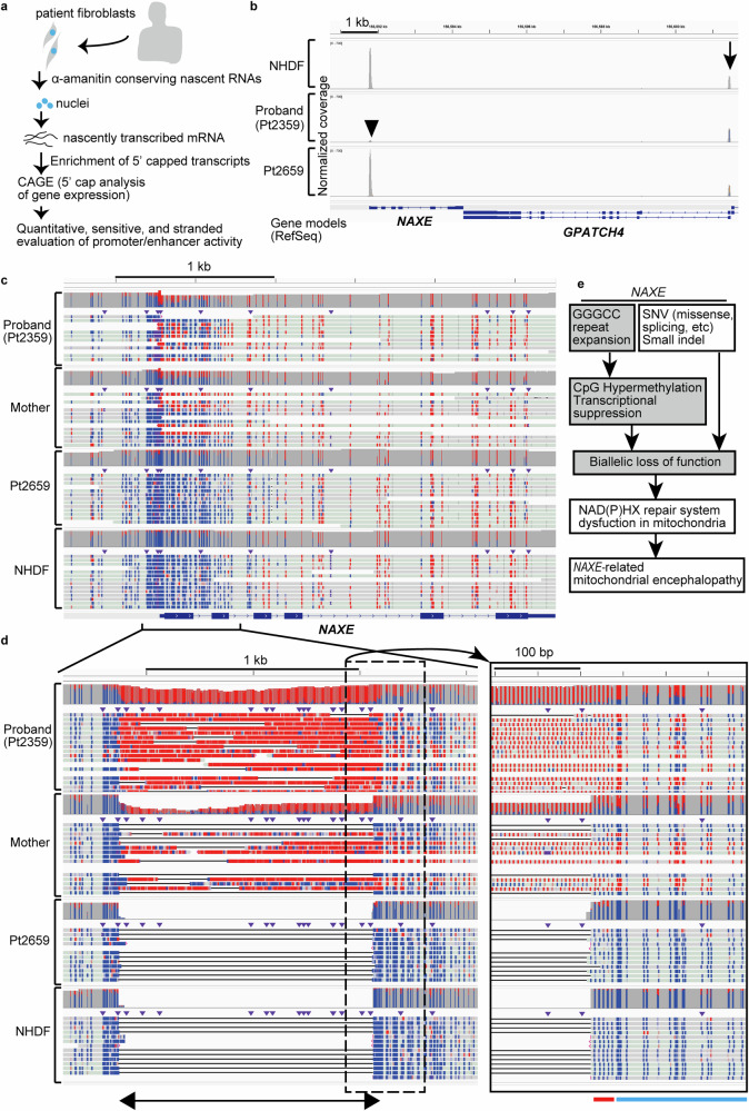
Repeat expansions cause at least 50 hereditary disorders, including Friedreich ataxia and other diseases known to cause mitochondrial dysfunction. We identified a patient with NAXE-related mitochondrial encephalopathy and novel biallelic GGGCC repeat expansion as long as ~200 repeats in the NAXE promoter region using long-read sequencing. In addition to a marked reduction in the RNA and protein, we found a marked reduction in nascent RNA in the promoter using native elongating transcript-cap analysis of gene expression (NET-CAGE), suggesting transcriptional suppression. Accordingly, CpG hypermethylation was observed in the repeat region. Genetic analyses determined that homozygosity in the patient was due to maternal chromosome 1 uniparental disomy (UPD). We assessed short variants within NAXE including the repeat region in the undiagnosed mitochondrial encephalopathy cohort of 242 patients. This study identified the GGGCC repeat expansion causing a mitochondrial disease and suggests that UPD could significantly contribute to homozygosity for rare repeat-expanded alleles.


Conflict of interest statement

The authors declare no competing interests.

Figures

Fig. 1
Fig. 1. Biallelic GGGCC repeat expansion in an NAXE-related mitochondrial encephalopathy patient.
a Our strategy for the identification of patients with outlying gene expression related to mitochondrial dysfunction. A total of 400 patients, including 303 patients with proven biochemical deficits, who were negative for pathogenic variants in the genetic screening, were analyzed by OUTRIDER. One of such OUTRIDER batches isolated a patient (Pt2359) with a marked decrease in NAXE expression. b “Family A” with markedly decreased NAXE expression in the proband. An arrow with “P” denotes the proband. -/-, +/-, and +/+ denote homozygote for wild type, heterozygote for repeat expansion, and homozygote, respectively. FLAIR MRI showed hyperintensity in brainstem and cerebellum (arrowhead). DWI and ADC showed restricted diffusion in part (arrows). c Volcano plot showing a remarkably decreased NAXE expression in the proband (arrow). d Western blotting showing loss of NAXE protein in the proband’s fibroblasts. NAXE positive controls (fHDF, fetal human dermal fibroblast (normal); C1 and C2, mitochondrial disease controls (Pt1615YS and Pt1753) who have no causative variants in NAXE and normal NAXE expression); Pt2659, another patient with a novel splicing variant and a missense variant; Beta-actin, a loading control. e Left panel, long-read sequencing of the proband showed the expansion of the GGGCC repeat (indicated as insertion (a dark violet tag with a number denoting inserted length of bases)) in each read (upper half) and the mother in a heterozygous state (lower half). Right panel, an example of such long-read sequence with rare inserts of slightly different repeat units (the red rectangle). f The site of repeat expansion with respect to NAXE gene body. g Waterfall plot showing repeat contents of Pt2359 and the mother for each long-read.
Fig. 2
Fig. 2. Genetic evaluation supporting the status of the NAXE locus in the pedigree.
a Design of repeat-primed polymerase chain reaction (RP-PCR) showing three primers (GGGCC_fwd primer and 5’ 6-FAM_reverse were used for initiating amplification between the repeat stretch and the adjacent genomic region, and RP_END primer was used for further efficient amplification) for the NAXE GGGCC repeat. b Segregation study using RP-PCR, showing Family A (left panel) and corresponding RP-PCR electropherogram detecting fluorescently labeled amplicons. Arrows in the mother and proband (Pt2359) indicate the presence of pathogenic GGGCC repeat expansion. c Probe design for southern blotting. Two small bars are probes that recognize the ~4.3 kb fragment created by MfeI and BamHI digestion of genomic DNA. (GGGCC)r denotes the GGGCC repeat in the NAXE promoter region. d Southern blotting confirmed the presence of a repeat expansion allele in the mother (I-2, in a heterozygous state) and proband (in a homozygous state). C, HapMap GM12878 sample with a normal range of repeat stretch as the control; M, molecular markers for southern blotting; arrow, expanded repeat allele; arrowhead, normal allele. e Homozygous stretch of single nucleotide variants (SNVs) using AutoMap on short read WGS data of the proband shows that most of the chromosome 1 regions were homozygous, suggesting uniparental disomy.
Fig. 3
Fig. 3. Amplicon long-read sequencing in the unidentified mitochondrial encephalopathy cohort.
a Design of amplicons for long-read sequencing. Two overlapping long-PCR amplicons covering the NAXE genomic region were designed. (GGGCC)r denotes the site of pathogenic GGGCC repeat expansion in the promoter region of NAXE. b Allele frequencies of variations in the GGGCC repeat region in the examined cohort and in TR-gnomAD, a dedicated tandem repeat catalogue, based on short-read whole genome sequencing (WGS). (GGGCC) ×5 denotes ×5 repeat and its derivatives. For details of each allele with different GGGCC repeat length or internal sequence, see Supplementary Table 4.
Fig. 4
Fig. 4. Functional consequence of GGGCC repeat expansions in NAXE.
a Experimental principles of NET-CAGE. The nuclei were isolated in the presence of α-amanitin, which maintains the nascently transcribed mRNAs. The nascent transcripts were enriched and processed using 5’ cap trapping technology to capture the full-length transcripts for subsequent sequencing (cap analysis of gene expression (CAGE)). b NET-CAGE of the proband fibroblasts compared to the controls (NHDF, normal neonatal human dermal fibroblast; Pt2659, patient with splicing and missense variants in NAXE) showing markedly suppressed nascent transcripts in the promoter region of NAXE (arrowhead). Note the comparable level of signals at GPATCH4 (arrow), which is reciprocally transcribed. c CpG hypermethylation (methylation indicated in red) was detected within the NAXE promoter region by analysis of long-read sequencing data in the proband as well as in the mother, whereas not in Pt2659 and NHDF. Reads were aligned to the reference genome (GRCh38). d Left panel, long-reads mapped against a sequence with expanded repeat showing hypermethylation in the GGGCC repeat per se as well as in the region downstream of the repeat stretch. The bidirectional arrow at the bottom denotes the repeat stretch. Right panel, magnified view of the dotted rectangle area in the left panel. Red and light blue lines below the right panel show the sites of normal length repeat in the controls (Pt2659 and NHDF) and the promoter region downstream of the repeat stretch, respectively. e Diagram of the suggested pathologic mechanism. GGGCC repeat expansion causes hypermethylation and suppressed transcription.

References

    1. DiMauro, S. & Schon, E. A. Mitochondrial respiratory-chain diseases. N. Engl. J. Med.348, 2656–2668 (2003). - PubMed
    1. Ng, Y. S. & Turnbull, D. M. Mitochondrial disease: genetics and management. J. Neurol.263, 179–191 (2016). - PMC - PubMed
    1. Skladal, D., Halliday, J. & Thorburn, D. R. Minimum birth prevalence of mitochondrial respiratory chain disorders in children. Brain126, 1905–1912 (2003). - PubMed
    1. Schlieben, L. D. & Prokisch, H. The Dimensions of Primary Mitochondrial Disorders. Front. Cell Dev. Biol.8, 600079 (2020). - PMC - PubMed
    1. Lynch, D. R. & Farmer, G. Mitochondrial and metabolic dysfunction in Friedreich ataxia: update on pathophysiological relevance and clinical interventions. Neuronal Signal.5, NS20200093 (2021). - PMC - PubMed

LinkOut - more resources