. 2024 Oct 25;14(1):25432.
doi: 10.1038/s41598-024-76269-1.

Development of a high-throughput screening system targeting the protein-protein interactions between PRL and CNNM

Affiliations

Development of a high-throughput screening system targeting the protein-protein interactions between PRL and CNNM

Yosuke Funato et al. Sci Rep. .

Abstract

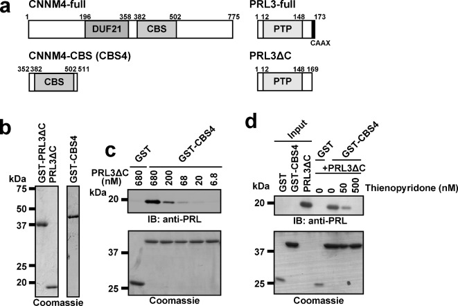
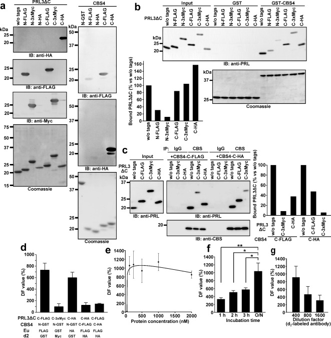
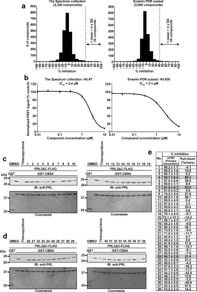
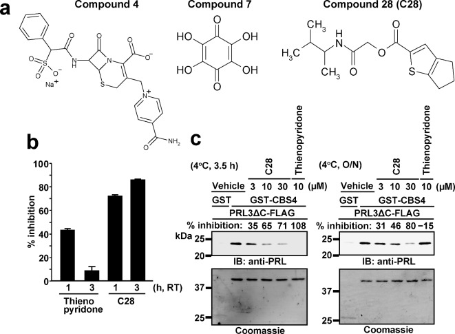
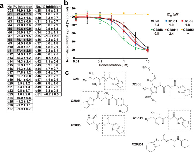
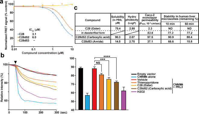
Phosphatase of regenerating liver (PRL) is an oncogenic protein that promotes tumor progression by directly binding to cyclin M (CNNM) membrane proteins and inhibiting their Mg2+ efflux activity. In this study, we have developed a high-throughput screening system to detect the interactions between PRL and CNNM proteins based on homogenous time-resolved fluorescence resonance energy transfer (HTR-FRET, HTRF). We optimized the tag sequences attached to the recombinant proteins of the CNNM4 CBS domains and PRL3 lacking the carboxyl terminal CAAX motif, and successfully detected the interaction by observing the FRET signal in the mixture of the tagged proteins and fluorophore-conjugated antibodies. Moreover, we performed compound library screening using this system and discovered several compounds that could efficiently inhibit the PRL-CNNM interaction. Characterization of one candidate compound revealed that it was relatively stable compared with thienopyridone, a known inhibitor of the PRL-CNNM interaction. The candidate compound can also inhibit PRL function in cells: suppression of CNNM-dependent Mg2+ efflux, and has sufficient in vitro drug metabolism and pharmacokinetic properties. Overall, these results demonstrate the effectiveness of this screening system for identifying novel inhibitors of the PRL-CNNM interaction, which could contribute to the development of novel anti-cancer drugs.

Keywords: Compound screening; Cyclin M (CNNM); HTRF); Homogenous time-resolved fluorescence resonance energy transfer (HTR-FRET; Phosphatase of regenerating liver (PRL); Protein-protein interaction (PPI).


Conflict of interest statement

The authors declare no competing interests.

Figures

Fig. 1
Fig. 1
Interaction between purified recombinant proteins of CNNM4-CBS domain and PRL3ΔC. (a) Schematics of the full length and deletion mutants of human CNNM4 and PRL3. The numbers of amino acid residues are indicated. DUF21: domain of unknown function 21, CBS: cystathionine β-synthase, PTP: protein tyrosine phosphatase. (b) Indicated purified recombinant proteins were separated by SDS-PAGE and subjected to Coomassie staining. (c, d) GST or GST-CBS4 proteins immobilized on beads were mixed with indicated concentrations of PRL3ΔC proteins (c), or 125 nM of PRL3ΔC proteins and indicated concentrations of thienopyridone (d). The proteins bound to the beads were subjected to immunoblotting with an anti-PRL antibody or Coomassie staining. Original blots/gels are presented in Supplementary Figure 1
Fig. 2
Fig. 2
Optimization of HTRF-based detection of PRL3ΔC-CBS4 interaction. (a) Purified recombinant PRL3ΔC (left) and CBS4 (right) proteins with indicated tags (N and C indicates the tag position) were separated by SDS-PAGE and subjected to immunoblotting with indicated antibodies or Coomassie staining. (b) GST or GST-CBS4 proteins immobilized on beads were mixed with indicated tagged PRL3ΔC proteins, and the proteins bound to the beads were subjected to immunoblotting with an anti-PRL antibody or Coomassie staining. The signal ratio of bound PRL3ΔC protein to their respective input is also indicated as a bar graph. (c) Indicated tagged PRL3ΔC and CBS4 proteins were mixed and subjected to immunoprecipitation with an anti-CBS antibody. The immunoprecipitates were subjected to immunoblotting with indicated antibodies or Coomassie staining. The signal ratio of bound PRL3ΔC protein to their respective input is also indicated as a bar graph. (d) Indicated tagged PRL3ΔC and CBS4 proteins (125 nM) were mixed with indicated Eu- and d2-conjugated antibodies (1/400 dilution) and incubated overnight at 4 oC, and subjected to HTRF analyses. The signal/background differences for each combination were indicated as DF value (in %, mean ± SD, n = 3). (e − g) PRL3ΔC-FLAG and GST-CBS4 proteins were subjected to HTRF analyses with varying (e) protein concentrations (mean ± SEM, n = 2 − 5), (f) incubation period (mean ± SEM, n = 5), and (g) dilution factors of d2-conjugated anti-GST antibody (mean ± SD, n = 3). *P < 0.05, **P < 0.01 by one-way ANOVA followed by Tukey’s test. Original blots/gels are presented in Supplementary Fig. 1.
Fig. 3
Fig. 3
Screening of compounds for PRL3ΔC-CBS4 PPI. (a) Primary screening results. Histograms of % inhibition of the normalized FRET signal from experiments with Spectrum collection (left) and Enamine PDR subset 2,000 (right) are shown. Dotted lines indicate the mean + 3 × SD threshold, and 58 compounds surpassed the threshold were determined as hit compounds. (b) As a secondary screening, each hit compound from the primary screening was mixed with PRL3ΔC-FLAG proteins at 7 different concentrations from 0.01 to 10 μM, and then subjected to HTRF analyses with GST-CBS4 proteins. Representative positive results showing the sigmoidal dose-dependency are shown with their IC50 values (mean ± SEM, n = 3). (c, d) The result of the tertiary screening. GST or GST-CBS4 proteins immobilized on beads were mixed with PRL3ΔC-FLAG proteins and indicated hit compounds (5 μM) from either Spectrum collection (c) or Enamine PDR subset 2,000 (d), and the proteins bound to the beads were subjected to immunoblotting with an anti-PRL antibody or Coomassie staining. (e) Summary of the compounds proceeded to the tertiary screening. The mean ± SD of % inhibition of normalized FRET signal in primary and secondary screening (at 10 μM, n = 2), and the % inhibition of coprecipitated PRL3ΔC-FLAG signal in tertiary screening, are indicated. Compounds with more than 90% mean inhibition in primary and secondary screening and more than 20% inhibition in tertiary screening are in grey backgrounds. Original blots/gels are presented in Supplementary Figure 1.
Fig. 4
Fig. 4
Comparison of compound 28 (C28) with thienopyridone. (a) Chemical structure of 3 candidate compounds selected from the screening results. (b) 10 μM of thienopyridone or C28 was mixed with PRL3ΔC-FLAG proteins, and then subjected to HTRF analyses after incubation for indicated time periods at room temperature with GST-CBS4 proteins and antibodies. % inhibition of each compound and incubation period are indicated as mean ± SEM (n = 2). (c) GST or GST-CBS4 proteins immobilized on beads were mixed with PRL3ΔC-FLAG proteins and indicated concentrations of thienopyridone or C28, and incubated for 3.5 h (left) or overnight (right) at 4 °C. The proteins bound to the beads were then subjected to immunoblotting with an anti-PRL antibody or Coomassie staining. % inhibition of each compound and incubation period was also indicated. Original blots/gels are presented in Supplementary Figure 1.
Fig. 5
Fig. 5
TR-FRET analyses for C28 derivatives. (a) C28 or each C28 derivative was mixed with PRL3ΔC-FLAG proteins at 10 μM, and then subjected to HTRF analyses with GST-CBS4 proteins. % inhibition values for each compound are indicated (mean ± SD, n = 3). Compounds that showed higher % inhibition than C28 (74.9%) are shown in a grey background and were selected for subsequent experiments. (b) Each of the 4 selected compounds in (a), C28, or C28d51 as a reference was mixed with PRL3ΔC-FLAG proteins at 7 different concentrations from 0.01 to 10 μM, and then subjected to HTRF analyses with GST-CBS4 proteins. The representative results with their IC50 values are shown (mean ± SEM, n = 3) (c) Chemical structures of C28 and derivative compounds used in (b). The dotted boxes indicate the 4H,5H,6H-cyclopenta[b]thiophene-2-carboxylic acid substructure. The chemical structures of all the derivative compounds are listed in Supplementary Table 1.
Fig. 6
Fig. 6
Characterization of carboxylic acid and amide derivatives of C28. (a) Each compound was mixed with PRL3ΔC-FLAG proteins at 7 different concentrations from 0.01 to 10 µM, and then subjected to HTRF analyses with GST-CBS4 proteins. The representative results with their IC50 values are shown (mean ± SEM, n = 3). (b) HEK293 cells transfected with the indicated constructs were loaded with Magnesium Green with high Mg2+ solution with compounds (30 µM) or H2O2 (200 µM), and then subjected to Mg2+-depletion at the indicated time point (arrowhead). The means of relative fluorescence intensities (left), and relative fluorescence intensities after Mg2+-depletion (at 5 min, mean ± SEM, n = 10 − 38) are indicated. ***P < 0.001, ****P < 0.0001 by one-way ANOVA followed by Tukey’s test. NS: not significant. (c) Solubility, Caco-2 monolayer permeability, and stability in human liver microsomes of indicated compounds were analyzed. Calculated hydrophobicity (logP) is also indicated. Italic values show the levels of deesterified form of C28, which was detected in the C28 samples. ND: not detected.

Similar articles

Cited by

References

    1. Diamond, R. H., Cressman, D. E., Laz, T. M., Abrams, C. S. & Taub, R. PRL-1, a unique nuclear protein tyrosine phosphatase, affects cell growth. Mol. Cell. Biol. 14, 3752–3762 (1994). - PMC - PubMed
    1. Saha, S. et al. A phosphatase associated with metastasis of colorectal cancer. Science 294, 1343–1346 (2001). - PubMed
    1. Bessette, D. C., Qiu, D. & Pallen, C. J. PRL PTPs: mediators and markers of cancer progression. Cancer Metastasis Rev. 27, 231–252 (2008). - PubMed
    1. Al-Aidaroos, A. Q. & Zeng, Q. PRL-3 phosphatase and cancer metastasis. J. Cell. Biochem. 111, 1087–1098 (2010). - PubMed
    1. Kozlov, G. et al. Structural insights into molecular function of the metastasis-associated phosphatase PRL-3. J. Biol. Chem. 279, 11882–11889 (2004). - PubMed

MeSH terms

LinkOut - more resources