. 2024 Oct 25;7(1):1387.
doi: 10.1038/s42003-024-07063-y.

A conserved Plasmodium nuclear protein is critical for late liver stage development

Affiliations

A conserved Plasmodium nuclear protein is critical for late liver stage development

Debashree Goswami et al. Commun Biol. .

Abstract

Malaria, caused by Plasmodium parasites, imposes a significant health burden and live-attenuated parasites are being pursued as vaccines. Here, we report on the creation of a genetically attenuated parasite by the deletion of Plasmodium LINUP, encoding a liver stage nuclear protein. In the rodent parasite Plasmodium yoelii, LINUP expression was restricted to liver stage nuclei after the onset of liver stage schizogony. Compared to wildtype P. yoelii, P. yoelii LINUP gene deletion parasites (linup-) exhibited no phenotype in blood stages and mosquito stages but suffered developmental arrest late in liver stage schizogony with a pronounced defect in exo-erythrocytic merozoite formation. This defect caused severe attenuation of the liver stage-to-blood stage transition and immunization of mice with linup - parasites conferred robust protection against infectious sporozoite challenge. LINUP gene deletion in the human parasite Plasmodium falciparum also caused a severe defect in late liver stage differentiation. Importantly, P. falciparum linup - liver stages completely failed to transition from the liver stage to a viable blood stage infection in a humanized mouse model. These results suggest that P. falciparum LINUP is an ideal target for late liver stage attenuation that can be incorporated into a late liver stage-arresting replication competent whole parasite vaccine.


Conflict of interest statement

Stefan Kappe, Ashley Vaughan, and Debashree Goswami are members of the Seattle Children’s Hospital workforce, and are inventors of patents licensed to Sanaria and have revenue distribution rights arising from patents rights licensed to Sanaria. All other authors declare no competing interests.

Figures

Fig. 1
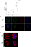
Fig. 1. P. yoeliiLiver stage Nuclear Protein (LINUP) (PY17X_1465200) localizes to the nucleus of liver stage parasites.
A Amino acid alignment between P. falciparum LINUP (PF3D7_1249700) and its syntenic orthologs P. yoelii LINUP (PY17X_1465200) and P. vivax LINUP (PVP01_1466800) shows 40% sequence identity and 60% sequence similarity. 80–100% sequence similarity is shown in red and 60 – 80% sequence similarity is shown in blue. The predicted and conserved nuclear localization sequence (NLS) is underlined in red and spans amino acids 133–156. B To visualize the localization of P. yoelii LINUP, an mCherry epitope-tagged parasite strain, P. yoelii LINUPmCherry was generated by fusing an mCherry tag to the LINUP C-terminus. The tagged transgenic parasite replaces the endogenous LINUP locus with the mCherry tagged LINUP. Immunofluorescence assays (IFAs) on P. yoelii LINUPmCherry infected mouse livers using an mCherry antibody (red), an antibody to the apicoplast acyl carrier protein (ACP) (green) and DAPI (blue) to delineate DNA, indicate that the protein is expressed at both 36 h and 48 h of infection and localizes to the nucleus of liver stage schizonts. Scale bar size is 20 μm. C At 36 h, LINUP (pink) shows partial co-localization with the acetylated histone H3 marker lysine 9 (green) and DAPI (white) with further magnification shown in D. Scale bar size is 10 μm for C and 2 μm for D.
Fig. 2
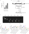
Fig. 2. P. yoelii linup creation and analysis of mosquito stage development.
A The schematic depicts the generation of the P. yoelii linup parasite using CRISPR/Cas9-mediated gene editing. Primers used to verify the gene deletion are indicated as P1, P2, P3 and P4 and the sizes of the PCR amplicons are shown in kilobases. B Agarose gel electrophoresis shows the PCR products corresponding to the gene deletion of P. yoelii LINUP in clones 3 (c3) and 5 (c5). C Blood stage growth was compared in groups of five Swiss Webster mice for P. yoelii XNL wildtype and P. yoelii linup clones c3 and c5. One million infected red blood cells were injected intravenously into each mouse and parasitemia was measured every other day for ten days. Growth rates were comparable suggesting LINUP has no essential role in blood stage replication. Data is represented as mean +/- SEM, n = 5 biological replicates. Statistical analysis was carried out using two-way ANOVA using Tukey’s multiple comparison test. P > 0.05 is taken as not significant. Source data is available as supplementary data. P. yoelii linup clones c3 and c5 did not have defects in mosquito infectivity as counts for (D), oocysts/midgut, (E), oocyst prevalence and (F), salivary gland sporozoites/mosquito were comparable between P. yoelii linup c3 (red) and c5 (green) to P. yoelii wildtype (black). Data is represented as mean +/- SD, n = 3 biological replicates. At least 20 mosquitoes were dissected for the counts shown in (D) and (E) and at least 100 mosquitoes were dissected for counts in (F). Statistical analysis was carried out using two-way ANOVA using Tukey’s multiple comparison test. P > 0.05 is taken as not significant.
Fig. 3
Fig. 3. Analysis of P. yoelii linup liver stage development.
Tissue sections were prepared from BALB/cJ mice infected with 250,000 sporozoites of either P. yoelii wildtype or P. yoelii linup from clone c3 at 24, 36 and 48 h post infection and analyzed by IFA. A Comparison of the size of liver stage parasites (based on area at the parasite’s largest circumference) between P. yoelii wildtype and P. yoelii linup at 24, 36 and 48 h indicate that P. yoelii wildtype liver stage schizonts are significantly larger than P. yoelii linup at 36 and 48 h. Data is represented as mean ± SD. Each datapoint refers to the mean size of at least 11 parasites for each timepoint. Statistical analysis was carried out using two-way ANOVA using Tukey’s multiple comparison test. P = 0.05 (*), ***P < 0.001, P > 0.05 is taken as not significant. B Liver stage development was compared at 48 h using antibodies against the parasite mitochondria (mHSP70, green) and apicoplast (acyl carrier protein, ACP, red) and in C, using antibodies to the parasite plasma membrane and mature exo-erythrocytic merozoite marker, MSP1 (red). DNA was stained with DAPI (blue). Scale bar: 10 μm. P. yoelii linup liver stage parasites are smaller compared to P. yoelii wildtype and display less branching of the mitochondria and apicoplast organelles. P. yoelii linup expresses the late liver stage proteins MSP1, however there is aberrant segregation of cytomeres and incomplete formation of mature exo-erythrocytic merozoites.
Fig. 4
Fig. 4. P. falciparum linup creation and analysis of mosquito stage development.
A Transcript per million (TPM) values are indicated for P. falciparum LINUP (PlasmoDB Identifier: PF3D7_1249700, NCBI Gene ID: 811529). P. falciparum LINUP gene expression steadily increases from day four to six of late liver stage development. RNA was isolated from P. falciparum infected primary hepatocytes (P. falciparum GFP+) from FRGN huHep mice on Day 2, 4 and 6 post infection. Data is represented as mean +/- SEM, of n = 3 biological replicates. B The schematic depicts the generation of P. falciparum linup knockout parasites using CRISPR/Cas9-mediated gene editing. Primers, P1, P2, P3 and P4 used to verify the gene deletion are indicated and the sizes of the PCR amplicons are shown in kilobases. C Agarose gel electrophoresis shows the PCR products corresponding to the gene deletion of P. falciparum linup. P. falciparum linup did not have defects in mosquito infectivity as counts for (D), oocysts/midgut, (E), oocyst prevalence and (F), salivary gland sporozoites/mosquito were comparable between P. falciparum linup clones B1 (blue), B4 (red), PIC4 (green), P2D7 (orange) and P. falciparum NF54 (black). Data is represented as mean +/- SD, of n = 2 biological replicates for P. falciparum linup clone B4 and n = 3 biological replicates for B1, P1C4 and P2D7. Statistical analysis was carried out using two-way ANOVA using Tukey’s multiple comparison test for (D) and one-way ANOVA using mixed-effects model (E) and (F). P > 0.05 is taken as not significant. At least 15 mosquitoes were dissected for analysis shown in (D) and (E) and at least 100 mosquitoes were dissected for the analysis shown in (F).
Fig. 5
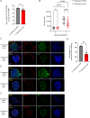
Fig. 5. P. falciparum linup liver stages display reduced growth and aberrant late liver stage development.
One million sporozoites from P. falciparum NF54 and P. falciparum linup clones B4 and P2D7 were injected four and seven FRG NOD huHep mice, respectively and livers were harvested on days five and seven. A Parasite liver load was quantified using 18S rRNA qRT-PCR on extracted RNA from infected livers on day seven. Data is represented as mean ± SD, n = 3 biological replicates for P. falciparum NF54 and n = 4 for P. falciparum linup. There is a statistically significant difference in liver load between P. falciparum NF54 and P. falciparum linup. Statistical analysis was carried out using unpaired t-test, P = 0.0388 (*). Tissue sections were prepared from FRG NOD huHep mice that were infected with one million sporozoites of P. falciparum NF54 and P. falciparum linup on days 5 and 7 post-sporozoite infection and analyzed for size and protein expression. B Comparison of the size of liver stage parasites (based on area at the parasite’s largest circumference) between P. falciparum NF54 and P. falciparum linup on days 5 and 7 post-sporozoite infection. No growth defect was seen in P. falciparum linup mid- liver stage schizonts on day five, however a significant growth defect was observed in late-liver stage schizonts on day seven. Data is represented as mean ± SD. Each datapoint refers to the mean size of at least 30 parasites for each timepoint. Statistical analysis was carried out using two-way ANOVA using Tukey’s multiple comparison test. ***P < 0.001, ****P < 0.0001, P > 0.05 is not significant (ns). C Left panel: IFA shows that at seven days of infection the P. falciparum NF54 parasitophorous vacuole membrane (PVM) protein EXP1 had a circumferential and contiguous staining pattern in the wildtype, as expected. In comparison EXP1 expression in mature P. falciparum linup day seven liver stages was patchy and aberrant. Expression of merozoite maker, MSP1, was weak and discontinuous in the P. falciparum linup. D Quantification of late liver stage schizonts with regard to a contiguous PVM based on the localization of the PVM marker EXP1 showed that ~65% of P. falciparum NF54 liver stages displayed a contiguous PVM while approximately only 30% P. falciparum linup displayed a contiguous EXP1 staining. Data is represented as mean ± SD, with n = 3 biological replicates for P. falciparum NF54 and n = 4 for P. falciparum linup. Statistical analysis was carried out using unpaired t-test. P = 0.0011 (**). E IFA shows that at seven days of infection the localization of parasite plasma membrane protein, CSP was visible in cytomeres for P. falciparum NF54, while for P. falicparum linup the distribution of CSP was aberrant. F IFA shows that at seven days of infection the parasite mitochondria protein HSP60 (mHSP60, red) and the apicoplast acyl carrier protein (ACP, green) in P. falciparum NF54 undergoes segregation in exo-erythrocytic stage merozoites, while these organelles remain branched in the P. falciparum linup liver stage parasite.
Fig. 6
Fig. 6. P. falciparum linup liver stages fail to transition to blood stages in FRG NOD huHep mice infused with human red blood cells.
A Schematic shows the experimental design of liver stage-to-blood stage transition experiments in FRG NOD huHep mice. One million sporozoites from P. falciparum NF54 and P. falciparum linup  from clones B1 and B4 were intravenuously injected into one, one and two  FRG NOD huHep mice, respectively. Mice were repopulated with human red blood cells as depicted. All mice were euthanized on day seven and 50 μl blood samples were collected from all mice for parasite load based on 18S rRNA qRT-PCR sampling. The remaining blood was transferred to in vitro culture and grown in culture for seven days. Further 50 μl blood samples were collected from in vitro culture samples for analysis of parasite 18S rRNA qRT-PCR samples seven days after transition to in vitro culture (14 days after sporozoite infection). B Analysis of parasite load by 18S rRNA qRT-PCR was carried out on extracted RNA from the blood of mice infected with P. falciparum NF54 (black bar) and P. falciparum linup (red bars) sporozoites on days 7 and day 14. On day 7, there was a severe defect in the P. falciparum linup transition; one of three mice infected with P. falciparum linup sporozoites was negative for 18S rRNA, while the other two infected mice had an approximate 10,000-fold reduction in parasite load as compared to wildtype. On day 14 after sporozoite infection wildtype NF54 blood stages significantly replicated during the intervening seven days gray bar, whilst no significant replication was seen for P. falciparum linup blood stages (red bars) suggesting that the liver stage-to-blood stage transition did not lead to viable blood stage parasites as also confirmed by lack of parasites detected in Giemsa-stained thin smears. C Schematic shows the experimental design of liver stage-to-blood stage transition experiments in FRG NOD huHep mice. One million sporozoites from P. falciparum NF54 and P. falciparum linup  clone P2D7 were injected into two and three FRG NOD huHep mice, respectively. Mice were repopulated with human red blood cells as depicted. All mice were euthanized on day seven and 50 μl blood samples were collected from all mice for parasite load based on 18S rRNA qRT-PCR sampling. The remaining blood was transferred to in vitro culture and grown in culture for three weeks. Further 50 μl blood samples were collected from in vitro culture samples for analysis of parasite 18S rRNA qRT-PCR samples on days 14, 21 and 28 after sporozoite infection and transition to in vitro culture. D Analysis of parasite load by 18S rRNA qRT-PCR was carried out on extracted RNA from the blood of mice infected with P. falciparum NF54 (gray bars) and P. falciparum linup (red bars) sporozoites. Both mice infected with P. falciparum NF54 (gray bars) sporozoites had ~1 × 106 parasites/ml seven days post infection, which had further increased to ~1 × 108 parasites/ml during the seven days of in vitro culture (day 14), and these were terminated on day 14. All three mice infected with P. falciparum linup (red bars) were negative for parasites by 18S rRNA qRT-PCR on all four timepoints (day 7, 14, 21, 28) indicating severe attenuation.

References

    1. World Health Organization. World Malaria Report 2022. World Health (WHO/HTM/GM, 2022).
    1. World Health Organization, W. WHO recommends groundbreaking malaria vaccine for children at risk. https://www.who.int/news/item/06-10-2021-who-recommends-groundbreaking-m... (2021).
    1. RTS, S. C. T. P. Efficacy and safety of RTS,S/AS01 malaria vaccine with or without a booster dose in infants and children in Africa: final results of a phase 3, individually randomised, controlled trial. Lancet386, 31–45 (2015). - PMC - PubMed
    1. Datoo, M. S. et al. Safety and efficacy of malaria vaccine candidate R21/Matrix-M in African children: a multicentre, double-blind, randomised, phase 3 trial. Lancet403, 533–544 (2024). - PubMed
    1. Seder, R. A. et al. Protection against malaria by intravenous immunization with a nonreplicating sporozoite vaccine. Science341, 1359–1365 (2013). - PubMed

Publication types

MeSH terms

LinkOut - more resources