. 2024 Oct 25;7(1):1390.
doi: 10.1038/s42003-024-07071-y.

Inflammation impacts androgen receptor signaling in basal prostate stem cells through interleukin 1 receptor antagonist

Affiliations

Inflammation impacts androgen receptor signaling in basal prostate stem cells through interleukin 1 receptor antagonist

Paula O Cooper et al. Commun Biol. .

Abstract

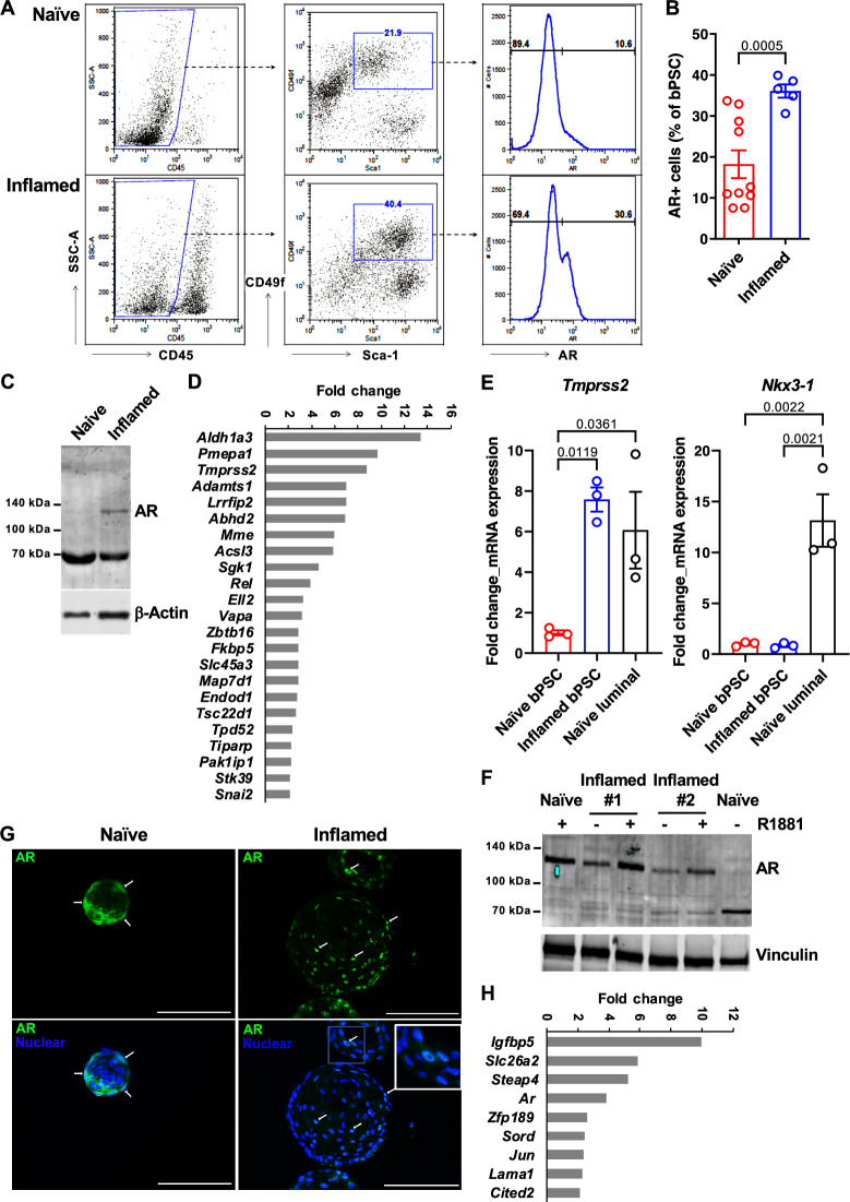
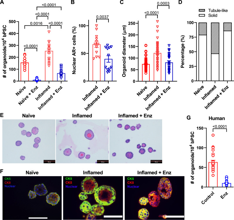
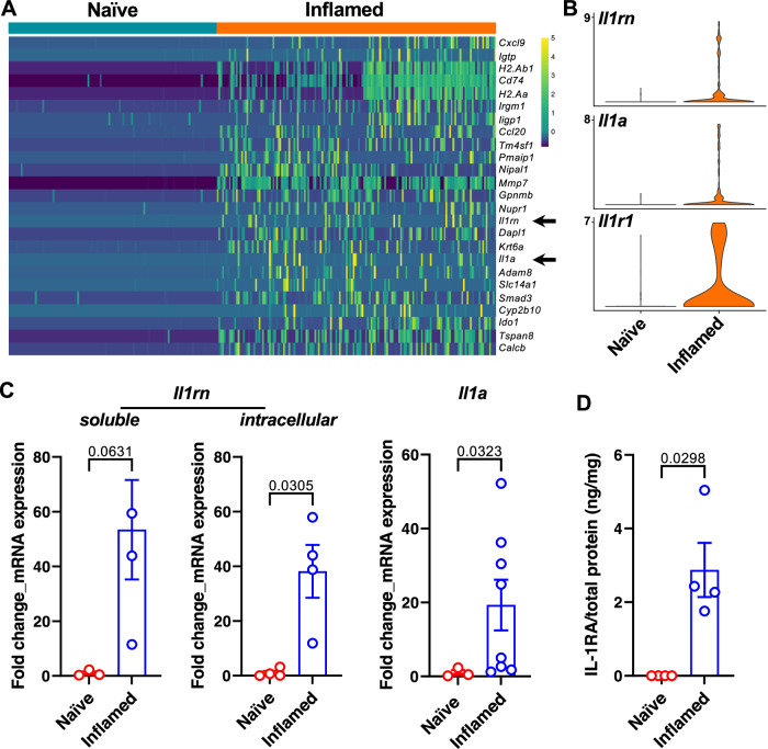
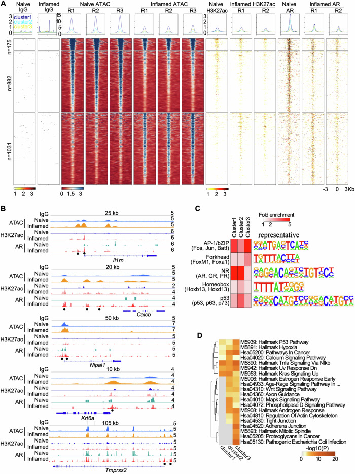
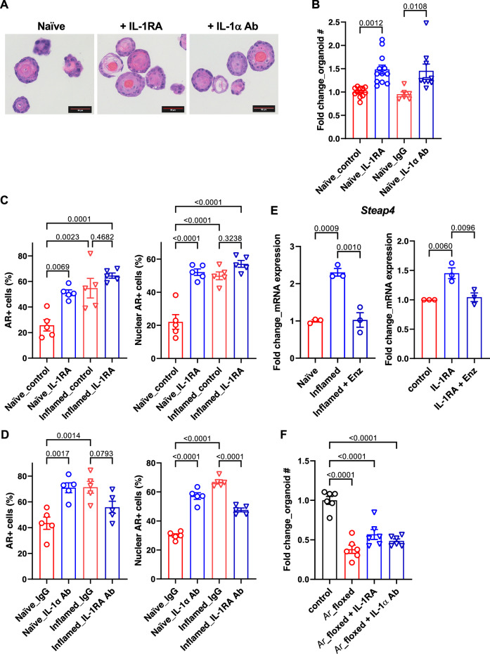
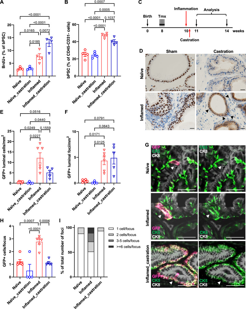
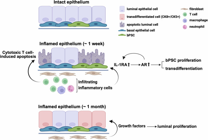
Chronic prostate inflammation in patients with benign prostate hyperplasia (BPH) correlates with the severity of symptoms. How inflammation contributes to prostate enlargement and/or BPH symptoms and the underlying mechanisms remain unclear. In this study, we utilize a unique transgenic mouse model that mimics chronic non-bacterial prostatitis in men and investigate the impact of inflammation on androgen receptor (AR) in basal prostate stem cells (bPSC) and their differentiation in vivo. We find that inflammation significantly enhances AR levels and activity in bPSC. More importantly, we identify interleukin 1 receptor antagonist (IL-1RA) as a crucial regulator of AR in bPSC during inflammation. IL-1RA is one of the top molecules upregulated by inflammation, and inhibiting IL-1RA reverses the enhanced AR activity in organoids derived from inflamed bPSC. Additionally, IL-1RA appears to activate AR by counteracting IL-1α's inhibitory effect. Furthermore, using a lineage tracing model, we observe that inflammation induces bPSC proliferation and differentiation into luminal cells even under castrate conditions, indicating that AR activation driven by inflammation is sufficient to promote bPSC proliferation and differentiation. Taken together, our study uncovers mechanisms through which inflammation modulates AR signaling in bPSC and induces bPSC luminal differentiation that may contribute to prostate hyperplasia.


Conflict of interest statement

The authors declare no competing interests.

Figures

Fig. 1
Fig. 1. AR expression and activity are elevated in bPSC freshly isolated from inflamed prostates and the enhanced AR activity is sustained in inflamed organoids in the absence of androgen.
A Representative flow cytometry dot plots and histograms of intracellular AR in bPSC from naïve and inflamed prostates. B Bar graph showing the percentage of AR+ cells within naïve and inflamed bPSC population as analyzed with flow cytometry (Naïve, n = 10; Inflamed, n = 5). C Immunoblot of full-length AR in naïve and inflamed bPSC lysates. D Fold change of AR target genes significantly upregulated in inflamed bPSC (Naïve or Inflamed, n = 3). E qRT-PCR of conventional AR target genes, Tmprss2 and Nkx3-1, in bPSC. Matched luminal population is included as a control (Naïve or Inflamed, n = 3). F Immunoblot showing increased level of full-length AR in inflamed organoids compared to naïve organoids. Full-length AR levels in both naïve and inflamed organoids were elevated with 1 nM R1881 treatment. G AR immunofluorescence staining in organoids derived from naïve or inflamed bPSC. Representative cells with nuclear-localized AR are indicated with white arrows. AR nuclear localization is also shown in the inset image with higher magnification (Scale bar, 100 μm). H Fold change of AR target genes significantly upregulated in inflamed organoids (Naïve or Inflamed, n = 3). Data presented in this figure are mean ± SEM.
Fig. 2
Fig. 2. Inhibition of AR activity reduces the differentiation of inflamed bPSC in organoid cultures.
A Number of organoids formed with naïve, inflamed bPSC with or without 10 μM Enz treatment (Naïve groups, n = 10; Inflamed groups, n = 18). B Quantitation of nuclear AR staining in inflamed organoids treated with Enz (n = 12). C Diameter of organoids and (D) percentage of organoids with tubule-like structures in naïve, inflamed organoids or inflamed organoids treated with Enz (Naïve, n = 74; Inflamed, n = 47; Inflamed+Enz, n = 25). E Representative H&E images of naïve, inflamed organoids or inflamed organoids treated with Enz (Scale bar, 100 μm). F CK5 (green) and CK8 (red) staining showing lack of organization in Enz-treated inflamed organoids (Scale bar, 25 μm). G Enz significantly reduces the number of organoids formed with human PSC (control or Enz, n = 27). Data presented in this figure are mean ± SEM.
Fig. 3
Fig. 3. IL-1RA is upregulated in inflamed bPSC.
A Il1rn and Il1a (arrows) are among the most upregulated genes in the inflamed bPSC compared to naïve bPSC as analyzed with scRNA-seq. B Violin plots of Il1rn, Il1a and Il1r1 expression in naïve and inflamed bPSC. C qRT-PCR of Il1rn isoforms and Il1a in naïve and inflamed bPSC (Naïve, n = 3–4; Inflamed, n = 3–8). D Upregulation of IL-1RA protein is confirmed using ELISA with lysates collected from naïve or inflamed bPSC (Naïve or Inflamed, n = 4). Data presented in this figure are mean ± SEM.
Fig. 4
Fig. 4. Genome-wide profiling of chromatin accessibility and AR binding.
A Histograms (above) and heatmaps (below) showing signal intensity of IgG, ATAC, H3K27ac, and AR peaks across naïve and inflamed bPSC, with biological replicates indicated as R1, R2, and R3. Differential peaks between naïve and inflamed bPSC with AR binding are displayed in three clusters. B Genome browser tracks at the Il1rn, Calcb, Nipal1, Krt6a, and Tmprss2 loci showing IgG, ATAC, H3K27ac, and AR signal in naïve and inflamed bPSC. Black dots represent peaks induced in inflamed bPSC. Track heights are indicated on the right corner for each track. C Enriched transcription factor (TF) DNA motifs at peak loci in each cluster. Shown are TF families on the left with representative TF members in parenthesis and a representative TF logo on the right. D Enrichment of KEGG and Hallmark pathways in genes adjacent to peaks from each cluster.
Fig. 5
Fig. 5. IL-1RA mediates inflammation-induced differentiation and AR activity in bPSC organoids.
A Representative H&E images of naïve organoids treated with recombinant IL-1RA or neutralizing antibody (Ab) against IL-1α, showing an increase in stratification of treated organoids (Scale bar, 50 μm). B Organoid formation with naïve bPSC was significantly increased with recombinant IL-1RA (50 ng/mL) or IL-1α neutralizing Ab treatment (5 μg/mL) (Naïve_control, n = 12; Naïve_IL-1RA, n = 12; Naïve_IgG, n = 6; Naïve_IL-1α Ab, n = 9). C Quantitation of AR+ (both overall and nuclear) cells in naïve or inflamed organoids treated with IL-1RA (50 ng/mL) (All groups, n = 5). D AR staining (overall and nuclear) was increased in naïve organoids treated with an IL-1α neutralizing Ab (5 μg/mL) and decreased in inflamed organoids treated with an IL-1RA neutralizing Ab (5 μg/mL) (All groups, n = 5). E qRT-PCR showing that the AR target gene Steap4, which was induced in inflamed organoids and suppressed with Enz (left, n = 3), was upregulated with IL-1RA in naïve organoids and Enz treatment abolished the upregulation (right, n = 3). F The effects of IL-1RA or IL-1α neutralizing Ab were lost with naïve organoids derived from Ar_flox/y bPSC which were deprived of AR (All groups, n = 6). Data presented in this figure are mean ± SEM.
Fig. 6
Fig. 6. Inflammation mimicking non-bacterial prostatitis promotes the proliferation of basal stem cells and the basal to luminal differentiation in vivo.
A Quantitation of BrdU+ bPSC in naïve and inflamed prostates, with or without castration (All groups, n = 4). B Percentage of bPSC in naïve and inflamed prostates, with or without castration (All groups, n = 4). C Experimental scheme showing time points of inflammation, castration and analysis. D AR staining shows maintenance of AR nuclear localization (arrow heads) in castrated inflamed prostates (Scale bar, 20 μm). E, F Quantitation of GFP+ cells or foci within the luminal layer (Naïve, n = 6; Naïve_castration, n = 4; Inflamed, n = 6; Inflamed_castration, n = 5). G Representative immunofluorescent images from naïve and inflamed prostates with or without castration showing the presence of GFP + CK5+ cells within the CK8+ luminal layer (arrowheads) (Scale bar, 50 μm). H, I Size of GFP+ foci and percentage of GFP+ foci of different sizes within the luminal layer (Naïve, n = 6; Naïve_castration, n = 4; Inflamed, n = 6; Inflamed_castration, n = 5). Data presented in this figure are mean ± SEM.
Fig. 7
Fig. 7. Diagram of enhanced AR signaling, bPSC proliferation, and differentiation that are mediated through IL-1RA during prostate inflammation that mimics non-bacterial prostatitis.
(Created in BioRender. Yang, J. (2023) BioRender.com/b35v181).

Update of

References

    1. Gandaglia, G. et al. The role of prostatic inflammation in the development and progression of benign and malignant diseases. Curr. Opin. Urol.27, 99–106 (2017). - PubMed
    1. De Nunzio, C., Presicce, F. & Tubaro, A. Inflammatory mediators in the development and progression of benign prostatic hyperplasia. Nat. Rev. Urol.13, 613–626 (2016). - PubMed
    1. Nickel, J. C. et al. The relationship between prostate inflammation and lower urinary tract symptoms: examination of baseline data from the REDUCE trial. Eur. Urol.54, 1379–1384 (2008). - PMC - PubMed
    1. Robert, G. et al. Inflammation in benign prostatic hyperplasia: a 282 patients’ immunohistochemical analysis. Prostate69, 1774–1780 (2009). - PMC - PubMed
    1. Lawson, D. A. et al. Basal epithelial stem cells are efficient targets for prostate cancer initiation. Proc. Natl. Acad. Sci. USA107, 2610–2615 (2010). - PMC - PubMed

Substances