Review
. 2024 Oct 25;22(1):655.
doi: 10.1186/s12951-024-02931-5.

The wonders of X-PDT: an advance route to cancer theranostics

Affiliations
Review

The wonders of X-PDT: an advance route to cancer theranostics

Asim Mushtaq et al. J Nanobiotechnology. .

Abstract

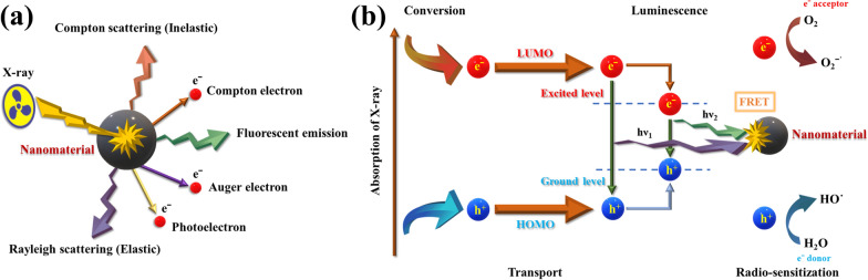
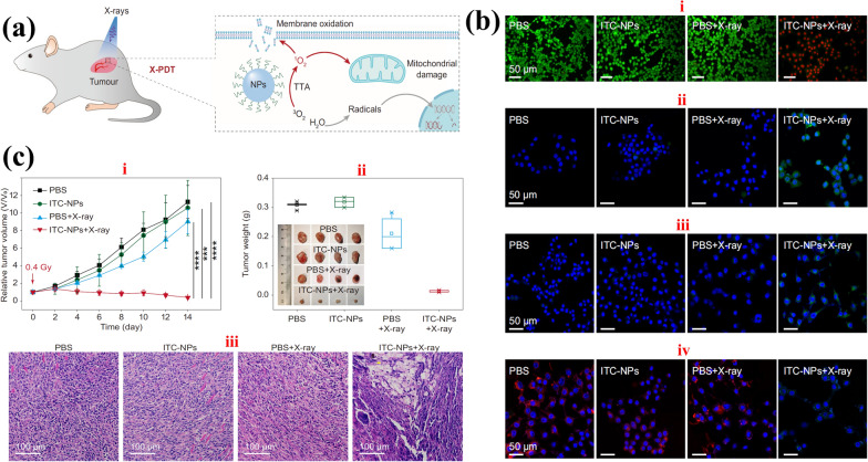
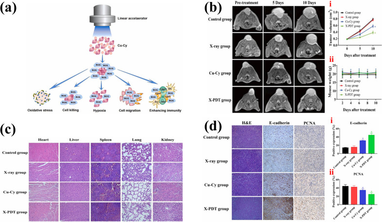
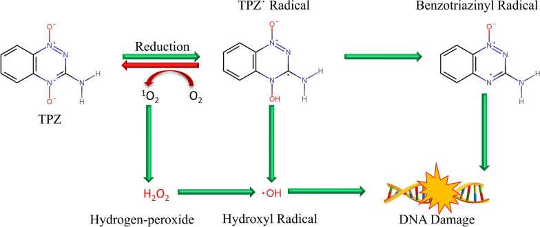
Global mortality data indicates cancer as the second-leading cause of death worldwide. Therefore, there's a pressing need to innovate effective treatments to address this significant medical and societal challenge. In recent years, X-ray-induced photodynamic therapy (X-PDT) has emerged as a promising advancement, revolutionizing traditional photodynamic therapy (PDT) for deeply entrenched malignancies by harnessing penetrating X-rays as external stimuli. Recent developments in X-ray photodynamic therapy have shown a trend toward minimizing radiation doses to remarkably low levels after the proof-of-concept demonstration. Early detection and real-time monitoring are crucial aspects of effective cancer treatment. Sophisticated X-ray imaging techniques have been enhanced by the introduction of X-ray luminescence nano-agents, alongside contrast nanomaterials based on X-ray attenuation. X-ray luminescence-based in vivo imaging offers excellent detection sensitivity and superior image quality in deep tissues at a reasonable cost, due to unhindered penetration and unimpeded auto-fluorescence of X-rays. This review emphasizes the significance of X-ray responsive theranostics, exploring their mechanism of action, feasibility, biocompatibility, and promising prospects in imaging-guided therapy for deep-seated tumors. Additionally, it discusses promising applications of X-PDT in treating breast cancer, liver cancer, lung cancer, skin cancer, and colorectal cancer.

Keywords: Deep tumors; ROS; Theranostic; X-PDT; X-ray responsive imaging.


Conflict of interest statement

The authors declare no competing interests.

Figures

Fig. 1
Fig. 1
Schematic representation of a various physical reactions that take place when X-rays come into contact with an X-ray responsive nanomaterial, b radio-sensitization caused by X-ray irradiation and a generic scintillation process
Fig. 2
Fig. 2
A graphical representation of applications of X-PDT against various types of cancer
Fig. 3
Fig. 3
a Schematic representation of in vivo therapy mechanism of deep seated tumor by X-PDT, b confocal laser scanning (CLSM) images of; (i) 4T1 cells co-staining analysis by Calcein-AM/PI, (live cells = green, dead cells = red); (ii) SOSG stained 4T1 cells; (iii) lipo-peroxides in 4T1 cells (ROS generation with BIODIPY C-11 staining is presented by green fluorescence); (iv) mitochondrial membrane potential of 4T1 cells (positive membrane potential = red fluorescence, decrease in membrane potential = green fluorescence), c representation of in vivo assessment of nano-scintillators; (i) 4T1 tumor growth curves; (ii) tumors weight after 14 days treatment; (iii) H&E-stained images of tumor slices. Reused with the permission of [74]. Copyright, The Author(s) 2022 under the license (http://creativecommons.org/licenses/by/4.0/)
Fig. 4
Fig. 4
a Schematic illustration of X-PDT influence on cancer cells, b MR-imaging for tumoricidal effect; (i) volume changes; (ii) body weight changes, c H & E staining of representative organs (kidney, heart, liver, lung and spleen), d immunohistochemistry (H & E, E-cadherin and PCNA staining of tumors) and morphological analysis; percentages positive expression of (i) E-cadherin; (ii) PCNA, (* p < 0.05 vs. control). Reused with the permission of [79]. Copyright, The Authors 2021, Publishing services by Elsevier B.V. under the license (http://creativecommons.org/licenses/by/4.0/)
Fig. 5
Fig. 5
A schematic diagram of cancer-killing under hypoxic environment by tirapazamine (TPZ)
Fig. 6
Fig. 6
a MRI of HCC in two different patients, first (i–iii) and second (iv–vi). Reused with the permission of [91]. Copyright, The Authors 2019, under the license (http://creativecommons.org/licenses/by/4.0/). b Transwell assays for the assessment of HepG2 and SK-Hep-1 migration, after the X-PDT with Cu-Cy NPs low dose (50 mg/L) as well as high dose (100 mg/L), c,d migratory cells counting (* p < 0.05 vs. control). Reused with the permission of [79]. Copyright, The Authors 2021, Publishing services by Elsevier B.V. under the license (http://creativecommons.org/licenses/by/4.0/)
Fig. 7
Fig. 7
Therapeutic outcomes in vivo. a BLI results of mice X-PDT with radiation dose of 5 Gy, b tumor growth analysis under BLI monitoring, c ex vivo BLI after dissection with left to right organs arrangement of top row: intestine, spleen, liver and skin; bottom row: muscle, brain, lung, heart and kidneys, d lungs BLI signal, based on ROI, e lung images of control and X-PDT groups, f H&E staining analysis (scale bar is 100 μm). Reused with the permission of [63]. Copyright, Ivyspring International Publisher under the terms and conditions (http://ivyspring.com/terms)
Fig. 8
Fig. 8
a A graphical presentation of synergistic therapy by Cu-Cy NPs, b images to present the morphology of B16 (melanoma) cells before and after X-ray irradiations and with and without Cu-Cy NPs treatment, respectively, c a presentation of cellular viability at different concentrations of Cu-Cy NPs with low dose (2.5 Gy) X-rays irradiation, d intracellular ROS generation by DCFH-DA assay, presenting increase in fluorescence intensity after exposure to X-rays, e,f measurement of rates of cell apoptosis and/or necrosis after treatment and irradiation, g tumor volumes measurement, h tumor growth curves, ik detection of infiltrative immune cells changes by flow cytometry. Reused with the permission of [49]. Copyright, The Authors 2020, under the license (http://creativecommons.org/licenses/by/4.0/)
Fig. 9
Fig. 9
a (i) A graphical presentation of targeted X-PDT; (ii) TEM image of FA-LPNPs-VP-5-FU, b confocal images of cellular viability of HCT116 cells (green fluorescence = dead cells); (i) quantitative analysis by ImageJ; (ii) calculated cellular viability by MTS assay after 24 h, c confocal images of apoptosis/necrosis assay; quantitative measurements of percentage changes at apoptosis/necrosis. Reproduced with the permission of [113]. Copyright, The Authors 2022, Published by Elsevier Masson SAS, under the license (https://creativecommons.org/licenses/by-nc-nd/4.0/)

Similar articles

Cited by

References

    1. Siegel RL, Miller KD, Fuchs HE, Jemal A. Cancer statistics, 2022. CA Cancer J Clin. 2022;72:7–33. - PubMed
    1. Mattiuzzi C, Lippi G. Current Cancer Epidemiology glossary. J Epidemiol Glob Health. 2019;9:217–22. - PMC - PubMed
    1. Debien V, De Caluwé A, Wang X, Piccart-Gebhart M, Tuohy VK, Romano E, et al. Immunotherapy in breast cancer: an overview of current strategies and perspectives. NPJ Breast Cancer. 2023;9:1–10. - PMC - PubMed
    1. Li X, Ramadori P, Pfister D, Seehawer M, Zender L, Heikenwalder M. The immunological and metabolic landscape in primary and metastatic liver cancer. Nat Rev Cancer. 2021;21:541–57. - PubMed
    1. Oudkerk M, Liu SY, Heuvelmans MA, Walter JE, Field JK. Lung cancer LDCT screening and mortality reduction—evidence, pitfalls and future perspectives. Nat Rev Clin Oncol. 2021;18:135–51. - PubMed

Substances

LinkOut - more resources