. 2024 Oct 25;17(1):256.
doi: 10.1186/s12920-024-02028-w.

RNA-seq validation of microRNA expression signatures for precision melanoma diagnosis and prognostic stratification

Affiliations

RNA-seq validation of microRNA expression signatures for precision melanoma diagnosis and prognostic stratification

Christopher G Love et al. BMC Med Genomics. .

Abstract

Background: New diagnostic tools are needed to improve the diagnosis and risk stratification of cutaneous melanoma. Disease-specific microRNA signatures have been previously described via NanoString profiling of solid biopsy tissue and plasma. This study validated these signatures via next-generation sequencing technology and compared their performance against clinical metrics and other published melanoma signatures.

Methods: RNA from 64 plasma and 60 FFPE biopsy samples from individuals with invasive melanoma or related benign/control phenotypes was extracted and enriched for microRNA. RNA sequencing was performed to compute MEL38/MEL12 signature scores. The results were evaluated with published NanoString and RNA sequencing datasets, comprising 548 solid tissue samples and 217 plasma samples, to predict disease status and patient outcome.

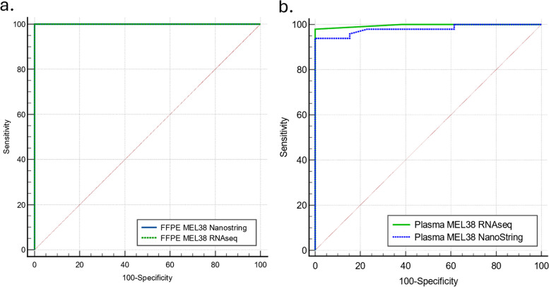
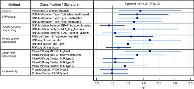
Results: The MEL38 diagnostic signature classifies patients into discrete diagnostic groups via RNA sequencing in either solid tissue or plasma (P < 0.001). In solid tissue, the prognostic MEL12 signature stratifies patients into low-, intermediate- and high-risk groups, independent of clinical covariates. The hazard ratios for 10-year overall survival, based on observed survival intervals, were 2.2 (MEL12 high-risk vs low-risk, P < 0.001) and 1.8 (intermediate-risk vs low-risk, P < 0.001), outperforming other published prognostic models. MEL12 also exhibited prognostic significance in the plasma of 42 patients with invasive disease.

Conclusions: The MEL38 and MEL12 signatures can be assessed in either solid tissue or plasma using RNA-seq and are strong predictors of disease state and patient outcome. Compared with other genomic methods, MEL12 was shown to be the strongest predictor of poor prognosis. MicroRNA expression profiling offers objective, accurate genomic information about a patient's likelihood of invasive melanoma and prognosis.

Keywords: Cancer; Gene expression; Melanoma; MicroRNA signatures; Precision medicine; RNA-seq.


Conflict of interest statement

CGL and RVL are employed and have equity in Geneseq Biosciences. LC is an employee of Australian Clinical Labs.

Figures

Fig. 1
Fig. 1
Schematic diagram of the study design. The diagram demonstrates the samples, datasets and technologies used to validate RNA-seq based microRNA profiling of plasma and solid tissue for melanoma diagnosis and prognosis
Fig. 2
Fig. 2
RNA-seq profiles from extracted total RNA demonstrating enrichment of microRNAs relative to other RNA species. a Macro-dissected FFPE tissue sections. b Plasma samples
Fig. 3
Fig. 3
MEL38 microRNA expression profiles of plasma samples and solid tissue samples grouped by melanoma stage. The relative differences in microRNA expression levels between clinically relevant disease states can be observed in both specimen types and analytical platforms. Dataset includes 59 plasma samples and 61 solid tissue samples. Red: high expression, Green: Low expression
Fig. 4
Fig. 4
ROC comparisons between methods and specimen types. a ROC curve comparison for 59 FFPE samples analysed with both RNA-seq and NanoString profiling. AUC for both methods: 1.0 (P < 0.001). b ROC comparison of MEL38 scores for 61 plasma samples profiled using both RNA-seq and NanoString. AUC for NanoString scores: 0.98 (P < 0.001), RNA-seq: 0.99 (P < 0.001). Pairwise comparison of curves; P = 0.32 (not significantly different)
Fig. 5
Fig. 5
Signature validation with public datasets. RNA sequencing (RNA-seq) data analysing MEL38 and MEL12 scores in 548 solid tissue and 217 plasma samples. a Box plot displaying MEL38 scores from RNA-seq analyses of solid tissue biopsies, categorized by disease status or melanoma stage. The dashed line depicts the MEL38 score threshold between invasive and non-invasive melanoma. b Kaplan–Meier survival analysis of melanoma patients grouped by solid tissue MEL12 scores (OBS). Log-rank test (P = 0.002). c Box plot showing MEL38 scores derived from RNA-seq analysis of plasma samples, grouped by disease status. d Kaplan–Meier survival analysis of invasive melanoma patients grouped by plasma MEL12 expression levels (MSS). Univariate Cox proportional hazards regression, P-value of 0.034. The log-rank test shows a P-value of 0.12
Fig. 6
Fig. 6
Comparative prognostic stratification of TCGA SCKM patients. Forest plot of Cox proportional hazard ratio values for the prediction of overall survival risk, using DNA, mRNA, microRNA sequencing or protein array-based classification of patients in the TCGA SKCM cohort (n = 354). Each CPH model included age, sex, stage and signature classification class. The keratin RNA-seq cluster (vs immune RNA-seq cluster) and the high-risk MEL12 signature (vs the low-risk group) had the highest hazard ratios (2.2, P < 0.001), reflecting their robust prognostic ability and independence of clinical covariates included in the analysis

References

    1. Fattore L, Costantini S, Malpicci D, Ruggiero CF, Ascierto PA, Croce CM, et al. MicroRNAs in melanoma development and resistance to target therapy. Oncotarget. 2017;8(13):22262–78. - PMC - PubMed
    1. Van Laar R, Lincoln M, Fereday S. Characterisation and validation of Mel38; A multi-tissue microRNA signature of cutaneous melanoma. PLoS One. 2019;14(2): e0211504. - PMC - PubMed
    1. Van Laar R, Lincoln M, Van Laar B. Development and validation of a plasma-based melanoma biomarker suitable for clinical use. Br J Cancer. 2018;118(6):857–66. - PMC - PubMed
    1. Stark MS, Tyagi S, Nancarrow DJ, Boyle GM, Cook AL, Whiteman DC, et al. Characterization of the Melanoma miRNAome by Deep Sequencing. PLoS One. 2010;5(3): e9685. - PMC - PubMed
    1. Van Laar R, Latif B, King S, Love C, Taubenheim N, Kalansooriya E, et al. Validation of a microRNA liquid biopsy assay for diagnosis and risk stratification of invasive cutaneous melanoma. Br J Dermatol. 2023;189(3):292–301. - PubMed

Publication types