. 2024 Sep 24;14(10):1203.
doi: 10.3390/biom14101203.

Insights into VDAC Gating: Room-Temperature X-ray Crystal Structure of mVDAC-1

Affiliations

Insights into VDAC Gating: Room-Temperature X-ray Crystal Structure of mVDAC-1

Kristofer R Gonzalez-DeWhitt et al. Biomolecules. .

Abstract

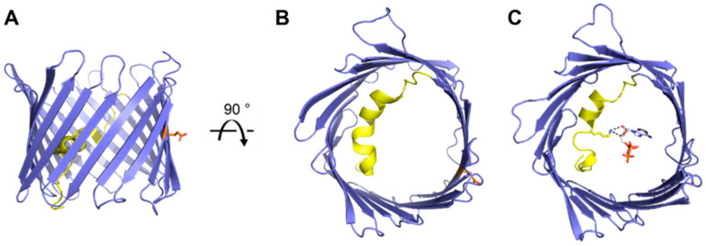
The voltage-dependent anion channel (VDAC) is a crucial mitochondrial protein that facilitates ion and metabolite exchange between mitochondria and the cytosol. Initially characterized over three decades ago, the structure of VDAC-1 was resolved in 2008, revealing a novel β-barrel protein architecture. This study presents the first room-temperature crystal structure of mouse VDAC-1 (mVDAC-1), which is a significant step toward understanding the channel's gating mechanism. The new structure, obtained at a 3.3 Å resolution, demonstrates notable differences from the previously determined cryogenic structure, particularly in the loop regions, which may be critical for the transition between the 'open' and 'closed' states of VDAC-1. Comparative analysis of the root-mean-square deviation (R.M.S.D.) and B-factors between the cryogenic and room-temperature structures suggests that these conformational differences, although subtle, are important for VDAC's functional transitions. The application of electric field-stimulated X-ray crystallography (EF-X) is proposed as a future direction to resolve the 'closed' state of VDAC-1 by inducing voltage-driven conformational changes in order to elucidate the dynamic gating mechanism of VDAC-1. Our findings have profound implications for understanding the molecular basis of VDAC's role in mitochondrial function and its regulation under physiological conditions.

Keywords: electric field-stimulated X-ray crystallography (EF-X); mitochondrial biology; room-temperature crystallography; voltage-dependent anion channel (VDAC).


Conflict of interest statement

The authors declare no conflict of interest.

Figures

Figure 1
Figure 1
Schematic representation of VDAC function in the cell. VDAC is located in the Outer Mitochondrial Membrane where it is the major pathway for exchange of metabolites and ions between the mitochondria and the cytosol. Strands 5–9 were omitted to show the interior of the barrel. IMS, intermembrane space. Prepared with Biorender.
Figure 2
Figure 2
Overview of VDAC-1 structures. Cartoon representation of mouse VDAC-1 (PDB 3EMN) as seen in the plane of the membrane (A) and from the intracellular side (B). Cartoon representation of mouse VDAC-1 with ATP bound (PDB 4C69) as seen from the intracellular side (C). The N-terminal helix is highlighted in yellow, sidechains for residues Lys-12 (yellow) and Glu-73 (orange) are shown as sticks representation, ATP is shown in grey, and hydrogen bonds between Lys-12 and ATP are shown in black.
Figure 3
Figure 3
Charge distribution on VDAC. Cartoon representation of mouse VDAC-1 (PDB 3EMN) as seen in the plane of the membrane (A) and from the intracellular side (B). Positively charged residues are highlighted in blue and negatively charged residues in red. Side change are shown as stick representation.
Figure 4
Figure 4
Overlay of PDB 3EMN (white) and the mVDAC-1 RT structure (teal) (A). Cartoon representation of mVDAC-1 RT as seen in the plane of the membrane (B) and from the intracellular side (C). The structure is colored according to the R.M.S.D. between mVDAC-1 RT and 3EMN. Regions with a high R.M.S.D. are shown in purple. The sidechain of residue Lys-12 is shown as a stick representation and the β-strands are numbered.
Figure 5
Figure 5
Temperature-dependent conformational change. 2Fo-Fc and Fo-Fo difference maps for loop regions with significant differences between the cryo and RT model (Fo-Fo difference map calculated by MatchMaps, correcting for limited isomorphism) (A,B). Maps are contoured to the indicated σ-levels, with all difference maps contoured the same. 2Fo-Fc maps were superimposed and are carved to within 2 Å of the model, and difference maps are carved to within 1.8 Å of the model. Red model: VDAC at 298 K. Blue model: VDAC at 100 K.
Figure 6
Figure 6
The beta barrel deforms slightly with temperature. (A) Side and top views of displacement vectors pointing from Cα positions in the cryo model to those in the room-temperature model. The β2 and β6 strands are colored gray. Only displacements larger than 0.5 Å are shown, magnified 5× for clarity. (B) 2Fo-Fc and Fo-Fo difference density for residues 94–103 of β6 and residues 38–47 of β2. Maps are contoured to the indicated σ-levels. 2Fo-Fc maps are carved to within 2 Å of the model, and difference maps are carved to within 1.8 Å of the model.
Figure 7
Figure 7
Overview of the mVDAC1 RT structure. The structure is colored according to the difference in the B-factor between the RT structure and PDB 3EMN. Regions with a small difference (indicating pre-existing high B-factors) are shown in purple. The residue Lys-12 sidechain is shown as a stick representation and β-strands are numbered. (A) View in the plane of the membrane. (B) View from the cytosolic side.

References

    1. Rostovtseva T., Colombini M. VDAC channels mediate and gate the flow of ATP: Implications for the regulation of mitochondrial function. Biophys. J. 1997;72:1954–1962. doi: 10.1016/S0006-3495(97)78841-6. - DOI - PMC - PubMed
    1. Varughese J.T., Buchanan S.K., Pitt A.S. The Role of Voltage-Dependent Anion Channel in Mitochondrial Dysfunction and Human Disease. Cells. 2021;10:1737. doi: 10.3390/cells10071737. - DOI - PMC - PubMed
    1. Chin H.S., Li M.X., Tan I.K.L., Ninnis R.L., Reljic B., Scicluna K., Dagley L.F., Sandow J.J., Kelly G.L., Samson A.L., et al. VDAC2 enables BAX to mediate apoptosis and limit tumor development. Nat. Commun. 2018;9:4976. doi: 10.1038/s41467-018-07309-4. - DOI - PMC - PubMed
    1. Shoshan-Barmatz V., Shteinfer-Kuzmine A., Verma A. VDAC1 at the Intersection of Cell Metabolism, Apoptosis, and Diseases. Biomolecules. 2020;10:1485. doi: 10.3390/biom10111485. - DOI - PMC - PubMed
    1. Ravi B., Kanwar P., Sanyal S.K., Bheri M., Pandey G.K. VDACs: An Outlook on Biochemical Regulation and Function in Animal and Plant Systems. Front. Physiol. 2021;12:683920. doi: 10.3389/fphys.2021.683920. - DOI - PMC - PubMed

Substances