. 2024 Oct 10;14(10):1274.
doi: 10.3390/biom14101274.

Postoperative Electroacupuncture Boosts Cognitive Function Recovery after Laparotomy in Mice

Affiliations

Postoperative Electroacupuncture Boosts Cognitive Function Recovery after Laparotomy in Mice

Yuen-Shan Ho et al. Biomolecules. .

Abstract

Postoperative cognitive dysfunction (POCD) is a common complication that affects memory, executive function, and processing speed postoperatively. The pathogenesis of POCD is linked to excessive neuroinflammation and pre-existing Alzheimer's disease (AD) pathology. Previous studies have shown that acupuncture improves cognition in the early phase of POCD. However, POCD can last for longer periods (up to weeks and years). The long-term effects of acupuncture are unknown. In this study, we hypothesized that electroacupuncture (EA) could reduce inflammation and cognitive dysfunction induced by laparotomy over a longer period. We characterized the effects of postoperative EA on cognitive changes and investigated the underlying molecular mechanisms in mice. Laparotomy was performed in 3-month-old mice followed by daily EA treatment for 2 weeks. Our data indicated that laparotomy induced prolonged impairment in memory and executive functions, which were mitigated by postoperative EA. EA also reduced tau phosphorylation and suppressed the activation of tau-related kinases and glia, with effects comparable to ibuprofen. These findings demonstrate the beneficial effects of EA in a mouse model of POCD, suggesting that EA's ability to suppress neuroinflammation may contribute to its protective effects. In conclusion, EA may be a viable non-pharmacological intervention for managing POCD in different phases of the medical condition.

Keywords: Chinese medicine; acupuncture; cognition; inflammation; memory; surgery; tau.


Conflict of interest statement

The authors declare no conflicts of interest.

Figures

Figure 1
Figure 1
Timeline of experimental procedures.
Figure 2
Figure 2
Electroacupuncture attenuated the cognitive impairment induced by laparotomy. The Y-maze test was used to assess associated memory at (a) 6 days (n = 8) and (d) 13 days (n = 14) after laparotomy. An increase in the number of errors made by the mice suggests cognitive impairment. The novel object recognition (NOR) test was used to assess hippocampal-dependent memory in the (b,c) early (n = 7) and (e,f) middle postoperative periods (n = 14–15). The data on days 5 and 12 in the NOR test showed that the mice had no preference among the two identical subjects placed in specific positions. The higher the discrimination index (TB/B+A), the better the novel object recognition ability of mice. Data were obtained from 2 to 3 batches of animals. Data were analyzed using one-way analysis of variance (ANOVA) followed by Tukey’s post hoc test for (a,f) and Kruskal–Wallis post hoc test for (c,d). * p < 0.05, ** p < 0.01, *** p < 0.001.
Figure 3
Figure 3
Electroacupuncture attenuated laparotomy-induced tau phosphorylation. Western blot analysis of changes in tau phosphorylation in the (a) hippocampus and (b) frontal cortex 14 days after laparotomy. Tau at different phosphorylation sites recognized by antibodies AT180 (pThr231/Ser235), AT8 (pSer202/Thr205), pTau404 (pSer404), and pTau396 (pSer396), and total tau data, were analyzed using one-way ANOVA followed by Tukey’s post hoc test for all the data. n = 7–8 for each group obtained from two batches of animals. * p < 0.05, ** p < 0.01, *** p < 0.001, **** p < 0.0001.
Figure 4
Figure 4
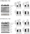
Electroacupuncture attenuated the activation of kinases. Western blot analysis of the expression of kinases in the (a) hippocampus and (b) frontal cortex in different groups 7 days after laparotomy. The image shows representative protein bands. Band intensities were measured using ImageJ, normalized to that of α-tubulin, and then analyzed using one-way ANOVA followed by Bonferroni’s post hoc test. n = 6–8 for each group. For all analyses, * p < 0.05, ** p < 0.01, *** p < 0.001, **** p < 0.0001.
Figure 5
Figure 5
Electroacupuncture had no effect on laparotomy-induced changes in synaptic proteins. Western blot analysis of the expression of synaptic proteins (synaptin-1, synaptophysin, and NMDAR2B) in the synaptosome fraction of the mouse (a) hippocampus and (b) frontal cortex 14 days after laparotomy. Representative protein bands are shown. Band intensities were measured using ImageJ, normalized to that of α-tubulin, and analyzed using one-way ANOVA followed by Tukey’s post hoc test. n = 5–7 for each group. For all analyses, * p < 0.05.
Figure 6
Figure 6
Levels of inflammatory cytokines after laparotomy. Neuroinflammation was investigated by detecting mRNA expression of pro-inflammatory cytokines in (a) the hippocampus and (b) the frontal cortex 7 days after laparotomy. n = 4–5. The mRNA expression levels of pro-inflammatory cytokines in (c) the hippocampus and (d) the frontal cortex 14 days after laparotomy. Data were analyzed using one-way ANOVA followed by Bonferroni’s post hoc test. n = 8–14. * p < 0.05, ** p < 0.01, *** p < 0.001.
Figure 7
Figure 7
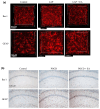
EA suppressed the activation of microglia and astrocytes after laparotomy. Immunohistochemical staining of brain slides for microglia (Iba-1) and astrocytes (GFAP) was performed using the (a) Accu-OPTICLear tissue-clearing technique. After all the behavioral tests, mice were sacrificed on day 14, and their brains were embedded for immunohistochemical staining using the transparent brain protocol, which allows better study of the 3D morphology of the target cells. Magnification = 40×. Scale bar = 50 μM. Images were taken from the CA1 region of the hippocampus for Iba-1-positive microglia and GFAP-positive astrocytes. Data represent 4 mice in each group. The results were confirmed using the (b) conventional ABC immunohistochemical staining method. Images were obtained from the hippocampal CA1 region. Data represent 3–4 mice (n = 3–4). Magnification = 10× Scale bar = 100 μM.
Figure 8
Figure 8
A comparison of cognitive performance between the EA and ibuprofen groups using Y-maze and NOR tests. The Y-maze test was used to assess associated memory (a) 6 days (n = 7) and (d) 13 days (n = 12) after laparotomy. The novel object recognition (NOR) test was used to assess hippocampal-dependent recognition memory in the (b,c) early (n = 7) and (e,f) middle period (n = 6–7) after laparotomy. Data were obtained from 2 to 3 batches of animals. Data were analyzed using an unpaired Student’s t-test. * p < 0.05, ** p < 0.01, ns = not significant.
Figure 9
Figure 9
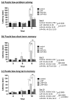
The investigation of executive function and cognitive ability in the middle postoperative period using the puzzle box test. An individual mouse was placed in a two-compartment arena. The experiments were conducted on days 10–13. The time taken to enter the goal zone was recorded. During this period, the entrance to the goal zone was blocked with different objects with increasing difficulty. Hence, the increased trial number in each figure represent the increased level of challenges to the mice. (a) Problem-solving ability was investigated in Trials 2, 5, and 8. (b) Short-term memory was investigated in Trials 3, 6, and 9. (c) Long-term memory was investigated in Trials 4, 7, and 10. Data were analyzed using two-way ANOVA followed by Turkey’s post hoc test. n = 6, * p < 0.05, ** p < 0.01, *** p < 0.001, **** p < 0.0001.

References

    1. Belrose J.C., Noppens R.R. Anesthesiology and cognitive impairment: A narrative review of current clinical literature. BMC Anesthesiol. 2019;19:241. doi: 10.1186/s12871-019-0903-7. - DOI - PMC - PubMed
    1. Vu T., Smith J.A. An Update on Postoperative Cognitive Dysfunction Following Cardiac Surgery. Front. Psychiatry. 2022;13:884907. doi: 10.3389/fpsyt.2022.884907. - DOI - PMC - PubMed
    1. Zhao Q., Wan H., Pan H., Xu Y. Postoperative cognitive dysfunction—Current research progress. Front. Behav. Neurosci. 2024;18:1328790. doi: 10.3389/fnbeh.2024.1328790. - DOI - PMC - PubMed
    1. Monk T.G., Weldon B.C., Garvan C.W., Dede D.E., van der Aa M.T., Heilman K.M., Gravenstein J.S. Predictors of cognitive dysfunction after major noncardiac surgery. Anesthesiology. 2008;108:18–30. doi: 10.1097/01.anes.0000296071.19434.1e. - DOI - PubMed
    1. Steinmetz J., Christensen K.B., Lund T., Lohse N., Rasmussen L.S. Long-term consequences of postoperative cognitive dysfunction. Anesthesiology. 2009;110:548–555. doi: 10.1097/ALN.0b013e318195b569. - DOI - PubMed

LinkOut - more resources