. 2024 Oct 12;14(10):1289.
doi: 10.3390/biom14101289.

Modulation of Carnitine Palmitoyl Transferase 1b Expression and Activity in Muscle Pathophysiology in Osteoarthritis and Osteoporosis

Affiliations

Modulation of Carnitine Palmitoyl Transferase 1b Expression and Activity in Muscle Pathophysiology in Osteoarthritis and Osteoporosis

Chiara Greggi et al. Biomolecules. .

Abstract

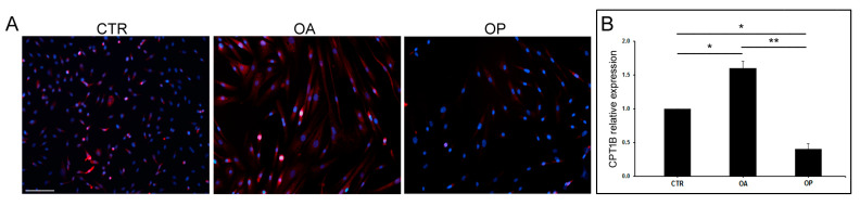
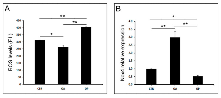
In the pathophysiology of osteoarthritis and osteoporosis, articular cartilage and bone represent the target tissues, respectively, but muscle is also involved. Since many changes in energy metabolism occur in muscle with aging, the aim of the present work was to investigate the involvement of carnitine palmitoyl transferase 1b (Cpt1b) in the muscle pathophysiology of the two diseases. Healthy subjects (CTR, n = 5), osteoarthritic (OA, n = 10), and osteoporotic (OP, n = 10) patients were enrolled. Gene expression analysis conducted on muscle and myoblasts showed up-regulation of CPT1B in OA patients; this result was confirmed by immunohistochemical and immunofluorescence analyses and enzyme activity assay, which showed increased Cpt1b activity in OA muscle. In addition, CPT1B expression resulted down-regulated in cultured OP myoblasts. Given the potential involvement of Cpt1b in the modulation of oxidative stress, we investigated ROS levels, which were found to be lower in OA myoblasts, and gene expression of nicotinamide adenine dinucleotide phosphate hydrogen oxidase 4 (Nox4), which resulted up-regulated in OA cells. Finally, the immunofluorescence of BCL2/adenovirus E1B 19 kDa protein-interacting protein 3 (Bnip3) showed a decreased expression in OP myoblasts, with respect to CTR and OA. Contextually, through an ultrastructural analysis conducted by Transmission Electron Microscopy (TEM), the presence of aberrant mitochondria was observed in OP muscle. This study highlights the potential role of Cpt1b in the regulation of muscle homeostasis in both osteoarthritis and osteoporosis, allowing for the expansion of the current knowledge of what are the molecular biological pathways involved in the regulation of muscle physiology in both diseases.

Keywords: energy metabolism; mitochondria biogenesis; muscle atrophy; myoblast regeneration; osteoarthritis; osteoporosis; oxidative stress.


Conflict of interest statement

The authors declare no conflicts of interest.

Figures

Figure 1
Figure 1
Morphological analyses and characterization of muscle fibers from CTR, OA, and OP patients. (AF) Representative images at different magnifications of hematoxylin–eosin-stained muscle sections from healthy (CTR), OA, and OP patients (scale bar = 100 µm). (GI) Immunohistochemical staining for myosin fast (scale bar = 100 µm). (J,K) Graphs showing morphometric evaluation of muscle fibers’ diameter and the percentage of type II muscle fibers in CTR, OA, and OP patients. Results are reported as mean ± SEM; t-test: * p < 0.05; ** p < 0.01.
Figure 2
Figure 2
CPT1B expression and activity in muscle tissue from CTR, OA, and OP patients. (A) Immunohistochemical staining for Cpt1b of muscle sections from healthy (CTR), OA, and OP patients (scale bar = 100 µm). (B) Graph showing the percentage of CPT1B-positive fibers in CTR, OA, and OP patients. (C) Cpt1b activity in muscle tissues from CTR, OA, and OP patients. (D) CPT1B transcript levels in muscle tissues from CTR, OA, and OP patients. Results are reported as mean ± SEM; t-test: * p < 0.05; ** p < 0.01. Abbreviation: O.D., optical density.
Figure 3
Figure 3
CPT1B expression in cultured myoblasts from CTR, OA, and OP patients. (A) Immunofluorescence for CPT1B in cultured myoblasts from healthy (CTR), OA, and OP patients (scale bar = 100 µm). (B) CPT1B transcript levels in cultured myoblasts from CTR, OA, and OP patients. Results are reported as mean ± SEM; t-test: * p < 0.05, ** p < 0.01.
Figure 4
Figure 4
Oxidative stress and NOX4 expression in cultured myoblasts from CTR, OA, and OP patients. (A) ROS levels in cultured myoblasts from healthy (CTR), OA, and OP patients. (B) NOX4 transcript levels in cultured myoblasts from CTR, OA, and OP patients. Results are reported as mean ± SEM; t-test: * p < 0.05; ** p < 0.01. Abbreviation: F.I., fluorescence intensity.
Figure 5
Figure 5
Bnip3 localization in cultured myoblasts from CTR, OA, and OP patients. Immunofluorescence for Bnip3 in cultured myoblasts from CTR, OA, and OP patients (scale bar = 100 µm). Specifically, in CTR myoblasts, the protein is diffuse along all the cytoplasm, whereas in OA myoblasts, Bnip3 is aggregated in numerous and distinct spots (puncta) similar to autophagosomes (white arrowheads). OP myoblasts revealed a lower expression of Bnip3 with no evident puncta.
Figure 6
Figure 6
Ultrastructural analysis of muscle tissue from CTR, OA, and OP patients. Transmission electron microscopy (TEM) of muscle tissue from CTR, OA, and OP patients. Images of CTR muscle fibers show the presence of well-organized sarcomeres (arrows), as well as for OA patients, while sarcomeres of OP muscle appear not well defined. Numerous glycogen granules (white arrowheads) characterize the muscle fibers of CTR and OA patients in contrast with OP group. Increased intermyofibrillar space is also observed in OP muscle, in which their presence appears reduced (double arrow). In addition, mitochondria (m) in the muscle tissue of CTR and OA patients display intact and well-organized cristae; in the muscle tissue of OP patients, on the contrary, mitochondria appeared irregularly shaped with loose mitochondrial cristae.

References

    1. Xie X., Fu J., Gou W., Qin Y., Wang D., Huang Z., Wang L., Li X. Potential Mechanism of Tea for Treating Osteoporosis, Osteoarthritis, and Rheumatoid Arthritis. Front. Med. 2024;11:1289777. doi: 10.3389/fmed.2024.1289777. - DOI - PMC - PubMed
    1. Piscitelli P., Neglia C., Feola M., Rizzo E., Argentiero A., Ascolese M., Rivezzi M., Rao C., Miani A., Distante A., et al. Updated Incidence and Costs of Hip Fractures in Elderly Italian Population. Aging Clin. Exp. Res. 2020;32:2587–2593. doi: 10.1007/s40520-020-01497-0. - DOI - PubMed
    1. Tarantino U., Celi M., Rao C., Feola M., Cerocchi I., Gasbarra E., Ferlosio A., Orlandi A. Hip Osteoarthritis and Osteoporosis: Clinical and Histomorphometric Considerations. Int. J. Endocrinol. 2014;2014:372021. doi: 10.1155/2014/372021. - DOI - PMC - PubMed
    1. Aibar-Almazán A., Voltes-Martínez A., Castellote-Caballero Y., Afanador-Restrepo D.F., Carcelén-Fraile M.D.C., López-Ruiz E. Current Status of the Diagnosis and Management of Osteoporosis. Int. J. Mol. Sci. 2022;23:9465. doi: 10.3390/ijms23169465. - DOI - PMC - PubMed
    1. Yokota S., Ishizu H., Miyazaki T., Takahashi D., Iwasaki N., Shimizu T. Osteoporosis, Osteoarthritis, and Subchondral Insufficiency Fracture: Recent Insights. Biomedicines. 2024;12:843. doi: 10.3390/biomedicines12040843. - DOI - PMC - PubMed

Substances

LinkOut - more resources