. 2024 Oct 19;14(10):1331.
doi: 10.3390/biom14101331.

A Novel Peptide from VP1 of EV-D68 Exhibits Broad-Spectrum Antiviral Activity Against Human Enteroviruses

Affiliations

A Novel Peptide from VP1 of EV-D68 Exhibits Broad-Spectrum Antiviral Activity Against Human Enteroviruses

Xiaojing Lin et al. Biomolecules. .

Abstract

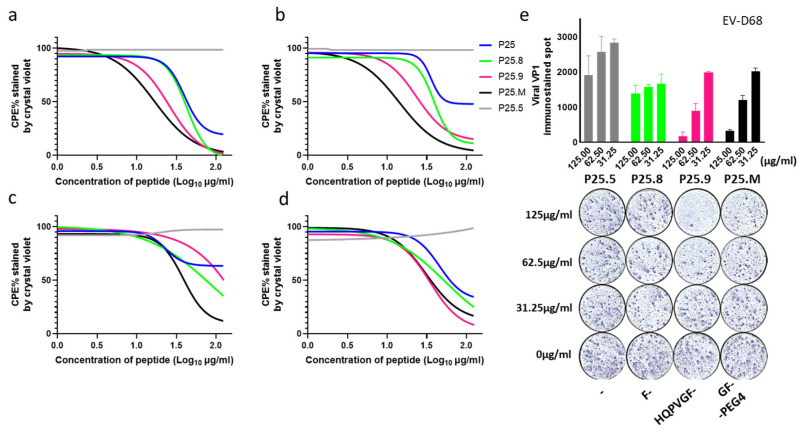
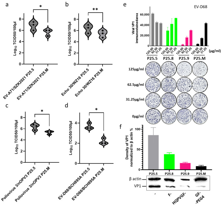
Enteroviruses have been a historical concern since the identification of polioviruses in humans. Wild polioviruses have almost been eliminated, while multiple species of non-polio enteroviruses and their variants co-circulate annually. To date, at least 116 types have been found in humans and are grouped into the species Enterovirus A-D and Rhinovirus A-C. However, there are few available antiviral drugs, especially with a universal pharmaceutical effect. Here, we demonstrate that peptide P25 from EV-D68 has broad antiviral activity against EV A-D enteroviruses in vitro. P25, derived from the HI loop and β-I sheet of VP1, operates through a conserved hydrophilic motif -R---K-K--K- and the hydrophobic F near the N-terminus. It could prevent viral infection of EV-A71 by competing for the heparan sulfate (HS) receptor, binding and stabilizing virions by suppressing the release of the viral genome. P25 also inhibited the generation of infectious viral particles by reducing viral protein synthesis. The molecular docking revealed that P25 might bind to the pocket opening area, a potential target for broad-spectrum antivirals. Our findings implicate the multiple antiviral effects of peptide P25, including blocking viral binding to the HS receptor, impeding viral genome release, and reducing progeny particles, which could be a novel universal anti-enterovirus drug candidate.

Keywords: EV-D68; antiviral; broad-spectrum; human enteroviruses; peptide.


Conflict of interest statement

The authors declare no conflicts of interest.

Figures

Figure 1
Figure 1
Peptide P11 and P25 exhibit antiviral potency to human enteroviruses. (a) Antiviral effects of peptide P1-P30 on EV-A71/FY0805, Echo 30/WZ16, and EV-D68/BCH895A. RD cells were infected with 100 TCID50/50 µL human enteroviruses co-incubated with 50 µL peptide P1-P30 at a concentration of 125 µg/mL (about 56 µM). 24 h post-infection, RD cells were stained using crystal violet and measured at 550 nm. The CPE was normalized to the only virus control (0%) and 0.5% DMSO mock (100%), and then converted to a white and blue heatmap. (b) The antiviral effects of the P25 homologous segments of VP1 from EV-A71 (P25.A71), Echo 30 (P25.E30), Poliovirus 3 (P25.PV3), Rhinovirus A81 (P25.A81), and Rhinovirus B70 (P25.B70). (c) Location of P11 and P25 at VP1 of EV-D68 (PDB: 6CRR). P11 and P25 are shown in yellow and magenta, respectively. (d) Peptide parameters of P11 and P25 calculated using the ProtParam tool. The positively charged amino acids were marked in red. P25 has a longer estimated half-life than P11. Sequences of P1-P30 are provided in Supplementary Table S1.
Figure 2
Figure 2
Antiviral activities of P25 mutants and its truncated peptides. (a) Sequences and parameters of the mutant P25s, parameters were calculated using the ProtParam tool. The positively charged amino acids were marked in red; “ + ” and “-” indicated the antiviral activity positive and negative, respectively; “ ++ ” indicated a broad spectrum of antiviral activity. The peptide P25.A81, was derived from Rhinovirus A81 and only inhibited EV-A71/FY0805 infection. The replacement of the N-terminal with G and F of P25.A81GF extended its antiviral profile. (b) RD cells were treated with 1 mg/mL of P25, P25.8, P25.9, P25.M, and P25.R, with serial 2-fold dilution for 24 h, and CC50 values were assayed using CCK8 reagents. P25.R was the peptide with a reverse sequence of P25 and had a stronger cytotoxic effect. So, the IC50 was not further tested as listed in Table 1. (cf) RD cells were infected with enteroviruses co-incubated with serial 2-fold diluted P25 mutants and its truncated peptides, stained using crystal violet and measured at 550 nm at 24 h post-infection. The CPE was normalized to the only virus control (0%) and a 0.5% DMSO mock (100%), and then converted to a white and blue heatmap. (c) Anti-EV-A71/FY0805 infection. (d) Anti-Echo 30/WZ16 infection. (e) Anti-Poliovirus 3/nOPV3 infection. (f) Anti-EV-D68/BCH895A infection.
Figure 3
Figure 3
P25 reduced the CPE of EV-A71/FY0805 via blocking viral binding to HS. (a) P25 at 62.5 µg/mL was used at the time points of pre-treatment (−6 h, −4 h, −2 h), co-treatment (0 h), and post-treatment (2 h, 4 h) with RD cells, and then the cells were infected with 100 TCID50/50 µL of EV-A71/FY0805. The cells were stained using crystal violet at 24 h post-infection. (b) The nonlinear curve fit of P25 pre-treatment with RD cells at −6 h, −4 h, −2 h compared with the co-treatment at 0 h. Pre-treatment enhanced the antiviral activity along with increased incubation time. The IC50 of P25 at 6 h of pre-treatment is 3.9 µg/mL. (c) Co-treatment of P25, P25.8, P25.9, and P25.M provides complete protection against 100 TCID50/50 µL of EV-A71/FY0805 with an IC50 of 32.35 ± 7.23 µg/mL, 24.49 ± 2.96 µg/mL, 13.02 ± 0.81 µg/mL, and 9.11 ± 2.52 µg/mL, respectively. P25.5 served as the negative control. (d) The antiviral activities of P25, P25.8, P25.9, and P25.M were interfered with by HS, which was a binding receptor for EV-A71/FY0805. Peptide P25s at a concentration of 62.5 µg/mL was pre-incubated with 500 µg/mL HS at 35°C for 1 h, then mixed with the virus and infected RD cells for 24 h. The cells were stained using crystal violet and measured at 550 nm. Data are presented as mean ± SD by three independent experiments. Statistical analysis was performed using a one-way ANOVA comparison. * indicates p < 0.05; ** indicates p < 0.01; *** indicates p < 0.001; ns indicates no statistical difference. (e,f) Antiviral performance of the P25s interrupted by serial diluted HS was shown using crystal violet and by the measurement at 550 nm. No significant interference was observed between HS and P25.5 with truncated G and F at the N-terminal, together with P25.11 removing the positive-charged amino acids. The findings suggested that positively charged amino acids were not the sole residues for binding with negatively charged HS.
Figure 4
Figure 4
Co-treatment of P25, P25.8, P25.9, and P25.M reduced CPE caused by human enteroviruses. P25, P25.8, P25.9, and P25.M co-treatment with enteroviruses from species A, B, C, and D at 35 °C for 1 h demonstrated anti-CPE effects. P25.8, P25.9, and P25.M have a broad spectrum of antiviral activity. P25.5 served as the negative control. Anti-CPE effects were nonlinear curve-fitted on four represented enteroviruses at 100 TCID50/50 µL. (a) EV-A71/SZK2021, which is not a HS-related strain. (b) Echo 30/WZ16. (c) Poliovirus 3/nOPV3. (d) EV-D68/BCH895A. Experiments were performed in triplicate. IC50 values are shown at Table 1. (e) 5 × 10⁴ RD cells were seeded 12 h before infection, infected with 10 TCID50/50 µL EV-D68 in the presence of P25.5, P25.8, P25.9, and P25.M at serial concentration for 1 h, and washed with DMEM twice, then replaced with DMEM for 24 h incubation. Viral VP1 protein was immune-stained and the stained focus was calculated using ImageJ (Version 1.54g).
Figure 5
Figure 5
P25s binding and thermostabilization of the virion. (a) EV-D68/BCH895A captured using a P25s coating ELISA and detected using anti-EV-D68 polyclonal Abs with a secondary antibody conjugated with HRP, binding affinities were compared by the fold increase normalized to the blank baseline (PBS). The procedure was described as in the Materials and Methods section. (be) The viral thermostabilization in the presence of P25, P25.5, P25.8, P25.9, and P25.M. About 4 µg of virus were mixed with 1.875 µg of P25, P25.5, P25.8, P25.9, and P25.M in 20 µL at 37 °C for 15 min and the temperature was subsequently increased to 90 °C, recording 10 points of the fluorescence signal at 1˚C intervals. The normalized genome release fluorescence dynamics and the first derivatives of EV-D68 (b,c) and Echo 30 (d,e) are shown. (f) The breakpoint temperature for genome release was calculated using the derivative of the fluorescence signal as the peak value. P25.8, P25.9, and P25.M increased the breakpoint temperature for the release of viral genome compared with P25.5 by approximately 5 °C for EV-D68/BCH895A and by 2–5 °C for Echo 30/WZ16. (g) P25.9 and P25.M retained the infectivity of Echo 30/WZ16. 106 TCID50/mL of Echo 30/WZ16 was co-incubated with an equal volume of the peptide at a concentration of 62.5 µg/mL at 37 °C for 15 min and 45 °C for 2 min, respectively, followed by rapid cooling on ice. The virus titer was determined using TCID50 as described in the Materials and Methods section. The experiment was repeated in triplicate. Statistical analysis was performed using paired two-tailed t-test. ** indicates p < 0.01.
Figure 6
Figure 6
P25 mutants docking to Echo 30 (PDB: 7C9S). Molecular docking analysis of P25s and the pentamer of Echo 30 using LeDock. The pocket opening of Echo 30 was marked with a black asterisk (*) and a single protomer was colored light blue to distinguish the other two neighboring protomers. The best scored pose was visualized using Pymol. (a) Pocket opening. (b) P25.9 (magenta). (c) P25.M (black). (d) Vacuum electrostatics on the surface of the pentamer with the peptide of P25.8 (green), P25.9 (magenta), and P25.M (black), where the pocket opening area has a higher potency for protein contact. Red indicates negative electrostatic potential energy; blue indicates positive electrostatic potential energy.
Figure 7
Figure 7
P25.M reduced the production of infectious virions. RD cells were infected with 100 TCID50 of enteroviruses for 1 h and washed using DMEM twice, then replaced with P25.M and P25.5 at a concentration of 62.5 µg/mL for a 24 h treatment, respectively. P25.5 served as the control. Data were presented in three independent experiments. Statistical analysis was performed using a paired two-tailed t-test. * indicates p < 0.05; ** indicates p < 0.01; (a) EV-A71/SZK2021. (b) Echo 30/WZ16. (c) Poliovirus 3/nOPV3. (d) EV-D68/BCH895A. (e) 5 × 10⁴ RD cells were seeded 12 h before infection, infected with 10 TCID50/50 µL of EV-D68 for 1 h and washed with DMEM twice, then replaced with P25.5, P25.8, P25.9, and P25.M at serial concentrations for a 24 h treatment. Viral VP1 was immune-stained and the stained area was calculated using ImageJ (Version 1.54g). P25.9 and P25.M at a concentration of 125 µg/mL, significantly inhibited the synthesis of the viral protein. (f) Western blotting for viral proteins. The infected cells in the presence of 62.5 µg/mL peptides were harvested, and the density of the viral protein band was calculated using ImageJ and normalized to β-actin as 100%. (The original image can be found in Supplementary File S1). The grey, green, pink and black color denotes the treatment with p25.5, p25.8, p25.9 and P25.M, respectively.

References

    1. Zhu P., Ji W., Li D., Li Z., Chen Y., Dai B., Han S., Chen S., Jin Y., Duan G. Current status of hand-foot-and-mouth disease. J. Biomed. Sci. 2023;30:15. doi: 10.1186/s12929-023-00908-4. - DOI - PMC - PubMed
    1. Imamura T. Enterovirus 68 among Children with Severe Acute Respiratory Infection, the Philippines. Emerg. Infect. Dis. 2011;17:1430–1435. doi: 10.3201/eid1708.101328. - DOI - PMC - PubMed
    1. Hsia S.-H., Lin J.-J., Chan O.-W., Lin T.-Y. Cardiopulmonary failure in children infected with Enterovirus A71. J. Biomed. Sci. 2020;27:53. doi: 10.1186/s12929-020-00650-1. - DOI - PMC - PubMed
    1. World Health Organization Poliomyelitis. [(accessed on 11 May 2024)]. Available online: https://www.who.int/news-room/fact-sheets/detail/poliomyelitis.
    1. Noisumdaeng P., Puthavathana P. Molecular evolutionary dynamics of enterovirus A71, coxsackievirus A16 and coxsackievirus A6 causing hand, foot and mouth disease in Thailand, 2000–2022. Sci. Rep. 2023;13:17359. doi: 10.1038/s41598-023-44644-z. - DOI - PMC - PubMed

MeSH terms

LinkOut - more resources