Review
. 2024 Sep 24;13(10):1156.
doi: 10.3390/antiox13101156.

Interplay between the Redox System and Renal Tubular Transport

Affiliations
Review

Interplay between the Redox System and Renal Tubular Transport

Xiao-Lan Wang et al. Antioxidants (Basel). .

Abstract

The kidney plays a critical role in maintaining the homeostasis of body fluid by filtration of metabolic wastes and reabsorption of nutrients. Due to the overload, a vast of energy is required through aerobic metabolism, which inevitably leads to the generation of reactive oxygen species (ROS) in the kidney. Under unstressed conditions, ROS are counteracted by antioxidant systems and maintained at low levels, which are involved in signal transduction and physiological processes. Accumulating evidence indicates that the reduction-oxidation (redox) system interacts with renal tubular transport. Redox imbalance or dysfunction of tubular transport leads to renal disease. Here, we discuss the ROS and antioxidant systems in the kidney and outline the metabolic dysfunction that is a common feature of renal disease. Importantly, we describe the key molecules involved in renal tubular transport and their relationship to the redox system and, finally, summarize the impact of their dysregulation on the pathogenesis and progression of acute and chronic kidney disease.

Keywords: kidney; metabolism; redox system; tubular transport.


Conflict of interest statement

The authors declare that they have no conflicts of interest.

Figures
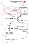
Figure 1
Figure 1
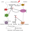
Schematic representation of the interplay between the redox system and mitochondrial metabolism. Mitochondria and the nicotinamide adenine dinucleotide phosphate oxidase (NOX) family are the major sources of endogenous ROS. Oxidative phosphorylation (OXPHOS) leads to the generation of ROS as metabolic by-products. Under pathological conditions, the interplay between lipid accumulation and mitochondrial dysfunction leads to ROS production. In turn, increased ROS lead to mitochondrial dysfunction and lipid accumulation. ROS play a critical role in Nuclear Factor kappa B (NF-κB), mitogen-activated protein kinases (MAPKs), phosphatidylinositol-3-kinase (PI3K)-Akt, and the KEAP1-NRF2-ARE signaling pathways. ROS are counteracted by antioxidants such as Nrf2 and GSH. Nrf2 upregulates target genes such as NAD(P)H quinone dehydrogenase 1 (NQO1), glutathione S-transferases (GSTs), catalase (CAT), and heme oxygenase 1 (HMOX), which play a critical role in mitigating oxidative stress, participating in detoxification pathways and increasing glutathione synthesis. The GSH pool is regulated by GPx and GST, which convert GSH to oxidized glutathione (GSSG); GR then reduces GSSG to GSH at the expense of NADPH, restoring the cellular GSH pool.
Figure 2
Figure 2
Schematic representation of key molecules involved in renal tubular transport and the redox system under physiological conditions. Under unstressed conditions, CD36 transports free fatty acids (FFAs) into the proximal tubular cells, which are then oxidized by fatty acid oxidation (FAO) and oxidative phosphorylation (OXPHOS) in the mitochondria. This process generates reactive oxygen species (ROS) as by-product. Organic anion transporter 1 (OAT1) and 3 (OAT3) mediate the uptake of uric acid from the blood into tubular cells, which exhibits a pro-oxidant behavior. MRP4 and breast cancer resistance protein (BCRP) are involved in the excretion of uric acid from tubular cells into the lumen of the nephron, thereby reducing intracellular levels of uric acid. Na+-K+-ATPase pumps 3 Na+ out of the cell and 2 K+ into the cell, creating an ion gradient across the cell membrane that drives the reabsorption of glucose and amino acids. Na+/H+ exchanger 3 (NHE3) reabsorbs HCO3 and about 80% of Na+ and also secretes H+. Glucose reabsorption depends mainly on sodium-glucose co-transporter 2 (SGLT2) and sodium-glucose co-transporter 1 (SGLT1). Glucose transporter 2 (GLUT2) mediates glucose transport across the basolateral membrane towards to the plasma. Glutamine is the most abundant amino acid in tubular cells and is reabsorbed through alanine-serine-cysteine transporter-2 (ASCT2) and BOAT1. Sodium-coupled neutral amino acid transporter-3 (SNAT3) and L-type amino acid transporter (LAT1) mediate the glutamine flux from blood to tubular cells. Once inside the cells, glutamine is converted to glutamate and used, along with cysteine and glycine, to produce GSH. GSH is regulated by the master regulator of redox balance, Nrf2, and Nrf2 transcriptional activity is inhibited by its main inhibitor, Keap1.
Figure 3
Figure 3
Schematic representation the interplay between key transporters and the redox system under stress conditions. Under stress conditions, CD36 expression is increased and transports more FFA into proximal tubular cells, leading to lipid accumulation and subsequent mitochondrial dysfunction and ROS production. In turn, increased ROS reduce mitochondrial lipid utilization, resulting in lipid accumulation and renal lipotoxicity. Increased ROS oxidize Na+-K+-ATPase subunits and promote Na+-K+-ATPase degradation, which may affect glucose and glutamine reabsorption. However, ROS activate Na+-K+-ATPase signaling pathways, which further increases the generation of mitochondrial ROS generation through the positive-feedback oxidant amplification loop. Oxidative stress inhibits the levels and activity of SGLTs and NHE3 in renal proximal tubule cells. Nrf2 is released from Keap1 and induces the expression of antioxidant genes. Nrf2 upregulates the expression of the glutamine transporter SNAT3 during metabolic acidosis. The effect of Nrf2 on SGLT2 is controversial. Overall, oxidative stress leads to renal inflammation, tubular apoptosis, and fibrosis and contributes to the development and progression of kidney disease. The lightning symbol indicates under stress conditions. The red arrows indicate increased expression, while the green arrows indicate decreased expression. Red “X” indicates prevention of the effect of Keap1 on Nrf2.

Similar articles

Cited by

References

    1. Lindeman R.D. Overview: Renal physiology and pathophysiology of aging. Am. J. Kidney Dis. 1990;16:275–282. doi: 10.1016/S0272-6386(12)80002-3. - DOI - PubMed
    1. Mandel L.J., Balaban R.S. Stoichiometry and coupling of active transport to oxidative metabolism in epithelial tissues. Am. J. Physiol. 1981;240:F357–F371. doi: 10.1152/ajprenal.1981.240.5.F357. - DOI - PubMed
    1. Imenez Silva P.H., Mohebbi N. Kidney metabolism and acid-base control: Back to the basics. Pflug. Arch. 2022;474:919–934. doi: 10.1007/s00424-022-02696-6. - DOI - PMC - PubMed
    1. Tian Z., Liang M. Renal metabolism and hypertension. Nat. Commun. 2021;12:963. doi: 10.1038/s41467-021-21301-5. - DOI - PMC - PubMed
    1. Le Gal K., Schmidt E.E., Sayin V.I. Cellular Redox Homeostasis. Antioxidants. 2021;10:1377. doi: 10.3390/antiox10091377. - DOI - PMC - PubMed

LinkOut - more resources