Review
. 2024 Oct 1;13(10):1193.
doi: 10.3390/antiox13101193.

Minimizing Oxidative Stress in the Lens: Alternative Measures for Elevating Glutathione in the Lens to Protect against Cataract

Affiliations
Review

Minimizing Oxidative Stress in the Lens: Alternative Measures for Elevating Glutathione in the Lens to Protect against Cataract

Julie C Lim et al. Antioxidants (Basel). .

Abstract

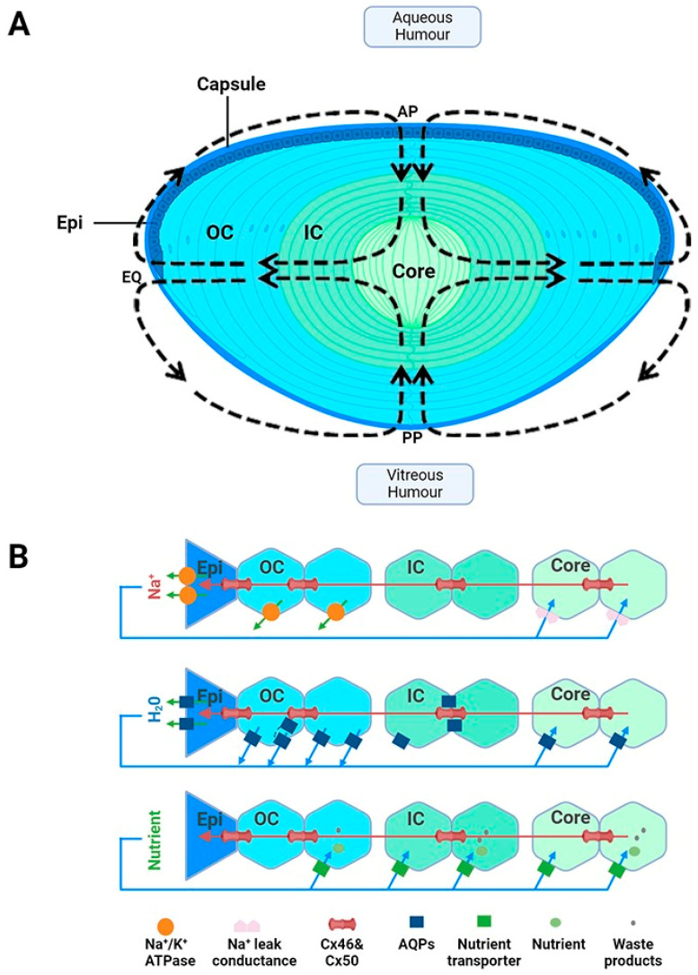
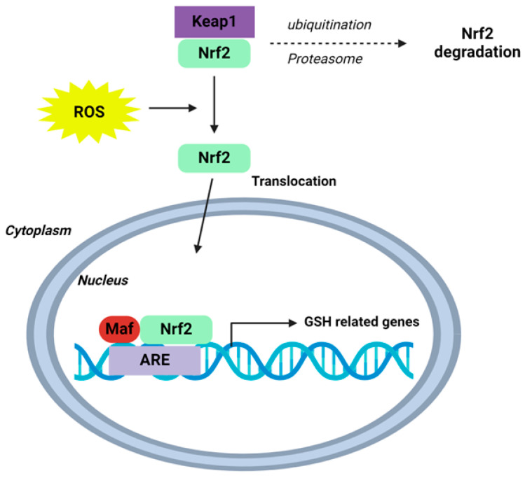
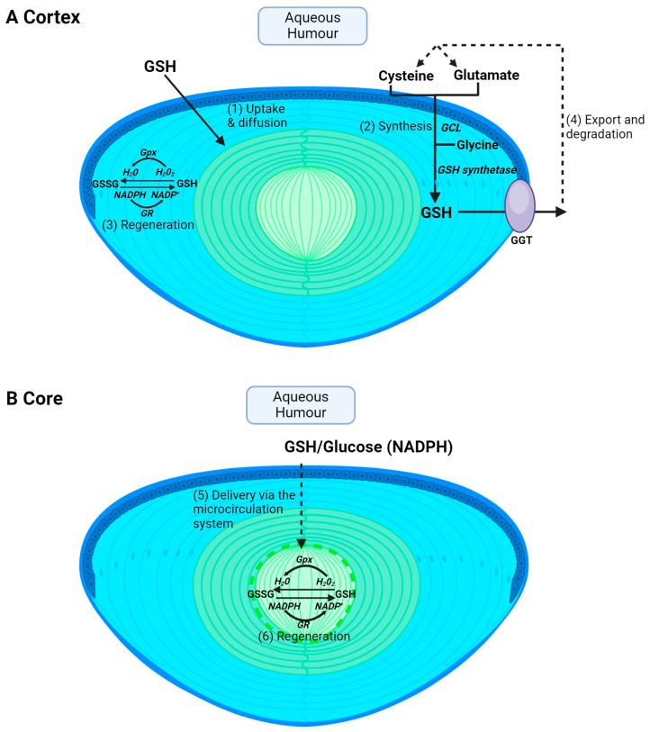
Oxidative stress plays a major role in the formation of the cataract that is the result of advancing age, diabetes or which follows vitrectomy surgery. Glutathione (GSH) is the principal antioxidant in the lens, and so supplementation with GSH would seem like an intuitive strategy to counteract oxidative stress there. However, the delivery of glutathione to the lens is fraught with difficulties, including the limited bioavailability of GSH caused by its rapid degradation, anatomical barriers of the anterior eye that result in insufficient delivery of GSH to the lens, and intracellular barriers within the lens that limit delivery of GSH to its different regions. Hence, more attention should be focused on alternative methods by which to enhance GSH levels in the lens. In this review, we focus on the following three strategies, which utilize the natural molecular machinery of the lens to enhance GSH and/or antioxidant potential in its different regions: the NRF2 pathway, which regulates the transcription of genes involved in GSH homeostasis; the use of lipid permeable cysteine-based analogues to increase the availability of cysteine for GSH synthesis; and the upregulation of the lens's internal microcirculation system, which is a circulating current of Na+ ions that drives water transport in the lens and with it the potential delivery of cysteine or GSH. The first two strategies have the potential to restore GSH levels in the epithelium and cortex, while the ability to harness the lens's internal microcirculation system offers the exciting potential to deliver and elevate antioxidant levels in its nucleus. This is an important distinction, as the damage phenotypes for age-related (nuclear) and diabetic (cortical) cataract indicate that antioxidant delivery must be targeted to different regions of the lens in order to alleviate oxidative stress. Given our increasing aging and diabetic populations it has become increasingly important to consider how the natural machinery of the lens can be utilized to restore GSH levels in its different regions and to afford protection from cataract.

Keywords: Nrf2; cataracts; cystine/cysteine; glutathione; lens microcirculation system.


Conflict of interest statement

J.C.L. and P.J.D. have collaborated with Nacuity Pharmaceuticals, Inc. in the testing of NACA and diNACA analogues on the lens.

Figures

Figure 1
Figure 1
Lens structure. (A): Architecture of the lens showing the capsule, anterior epithelial monolayer (Epi), elongating nucleated outer cortical fibre cells (OC), anucleated inner cortical fibre cells (IC) and mature fibre cells in the lens core (C). Fibre cells from adjacent hemispheres meet at the anterior (AP) and posterior (PP) poles to form the sutures. Arrows in the diagram represent the direction of ion and water fluxes that underpin the lens microcirculation system. (B): Top panel—an equatorial cross section of the lens showing a cellular view of ion movement in the lens. Current and solutes are proposed to flow into the lens via the extracellular space, to cross fibre cell membranes, and to flow outward via a gap-junction-mediated pathway (Cx 56 and Cx 50) at the lens equator (EQ) where the Na+/K ATPase pumps are concentrated. Middle panel—solute movement results in water crossing the membranes via aquaporin (AQP) water channels, which possess different water permeabilities and exhibit regional differences in their expression, subcellular localization, and regulation, thereby differentially contributing to water influx, outflow, and efflux, respectively. Bottom panel—isotonic fluid flux delivers nutrients to the lens nucleus, where they are accumulated by nutrient uptake transporters. Unwanted metabolites or waste products can then be removed from the lens. Adapted from [37] with permission from Elsevier.
Figure 3
Figure 3
Schematic of the Nrf2-Keap1-ARE signalling pathway. Under normal conditions, nuclear erythroid-2 like factor-2 (Nrf2) is ubiquitinated through Kelch-like ECH-associated protein1 (Keap1) and degraded in the proteasome. After ROS exposure, Keap1 is inactivated and Nrf2 translocates to the nucleus and binds to antioxidant response element (ARE) sites, subsequently activating many genes, including those involved in GSH homeostasis. Schematic adapted from [118]. Creative Commons Licence CCBY 4.0.
Figure 2
Figure 2
Regional differences in GSH accumulation in the lens. The different structural adaptations of the lens mean that the cortex and core accumulate GSH via different pathways. (A) In the cortex, GSH can be taken up directly from the aqueous humour via GSH transporters and then GSH can be supplied to cortical fibres via diffusion through gap junctions (1). Within cortical fibre cells, GSH can also be synthesised intracellularly from precursor amino acids, cysteine, glutamate, and glycine, by the sequential actions of the enzymes glutamate cysteine ligase (GCL) and glutathione synthetase (2). GSH can also be regenerated from GSSG. GSH is used as a co-factor for glutathione peroxidase (Gpx) in the process of detoxifying H2O2 into H2O and is in turn oxidised to GSSG. Glutathione reductase (GR) is used in combination with NADPH to help recycle GSSG back to GSH (3). Finally, GSH can be exported from the lens followed by degradation into its precursor amino acid by GGT and subsequent reuptake of amino acids for GSH synthesis (4). (B) In the core, passive diffusion is insufficient to deliver GSH to the lens’s centre. Instead, the microcirculation delivers GSH to the core where it can then be taken up into cells via GSH transporters (5). GSH levels can also be maintained by GSSG regeneration (6) but cannot be synthesised in the core.
Figure 4
Figure 4
Regulation of the microcirculation system. Lens water transport (pressure) is regulated by a dual-feedback pathway. Lens surface pressure (Pset) is maintained by two arms of a dual-feedback system that regulates the ion transporters that control the intracellular osmolarity of cells at the lens surface. Increases in pressure (ΔPi) due to hypoosmotic stress or increased zonular tension work via TRPV4 to activate a signalling pathway that involves the release of ATP via hemichannels, the subsequent activation of purinergic P2Y receptors, and prompts the Src family of protein tyrosine kinases (SFK) to increase the activity of the Na+/K+-ATPase and decrease lens pressure. Decreases in pressure (ΔPi), due to hyperosmotic stress or decreased zonular tension, work via TRPV1 to activate the extracellular signal-regulated kinase 1/2 (ERK1/2), phosphatidylinositol 3-kinase (PI3K)/Akt, kinase with no lysine (WNK), and Ste20-related proline–alanine-rich kinase (SPAK)/oxidative stress-responsive kinase-1 (OSR1) signalling pathways to directly activate the sodium potassium dichloride cotransporter (NKCC) and reduce the activity of the Na+/K+-ATPase to increase lens pressure. Adapted from [37] with permission from Elsevier.

Similar articles

Cited by

References

    1. Brian G., Taylor H. Cataract blindness—Challenges for the 21st century. Bull. World Health Organ. 2001;79:249–256. - PMC - PubMed
    1. Pascolini D., Mariotti S.P. Global estimates of visual impairment: 2010. Br. J. Ophthalmol. 2012;96:614–618. doi: 10.1136/bjophthalmol-2011-300539. - DOI - PubMed
    1. Age-Related Eye Disease Study Research G. A randomized, placebo-controlled, clinical trial of high-dose supplementation with vitamins C and E and beta carotene for age-related cataract and vision loss: AREDS report no. 9. Arch. Ophthalmol. 2001;119:1439–1452. doi: 10.1001/archopht.119.10.1439. - DOI - PMC - PubMed
    1. Mathew M.C., Ervin A.M., Tao J., Davis R.M. Antioxidant vitamin supplementation for preventing and slowing the progression of age-related cataract. Cochrane Database Syst. Rev. 2012;13:CD004567. doi: 10.1002/14651858.CD004567.pub2. - DOI - PMC - PubMed
    1. Bouayed J., Bohn T. Exogenous antioxidants--Double-edged swords in cellular redox state: Health beneficial effects at physiologic doses versus deleterious effects at high doses. Oxidative Med. Cell. Longev. 2010;3:228–237. doi: 10.4161/oxim.3.4.12858. - DOI - PMC - PubMed

LinkOut - more resources