. 2024 Oct 15;13(10):1237.
doi: 10.3390/antiox13101237.

Frequency-Dependent Antioxidant Responses in HT-1080 Human Fibrosarcoma Cells Exposed to Weak Radio Frequency Fields

Affiliations

Frequency-Dependent Antioxidant Responses in HT-1080 Human Fibrosarcoma Cells Exposed to Weak Radio Frequency Fields

Hakki Gurhan et al. Antioxidants (Basel). .

Abstract

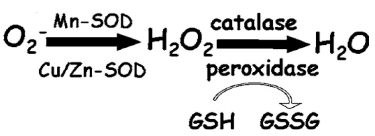
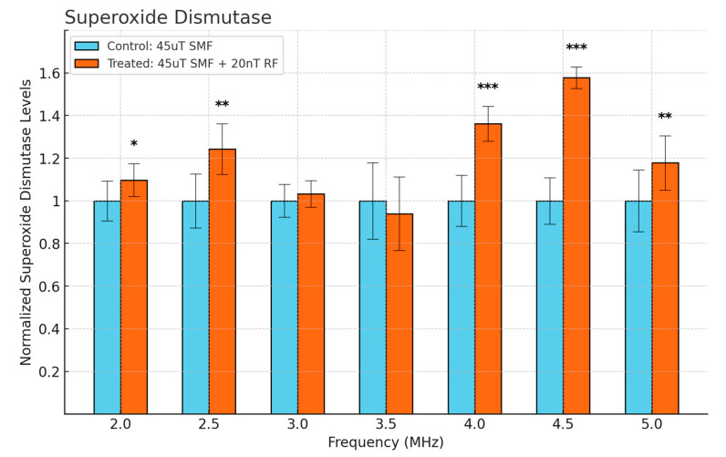
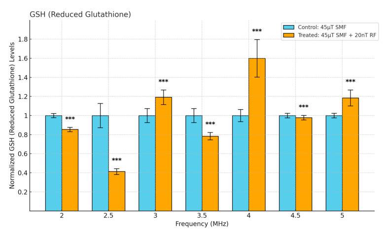
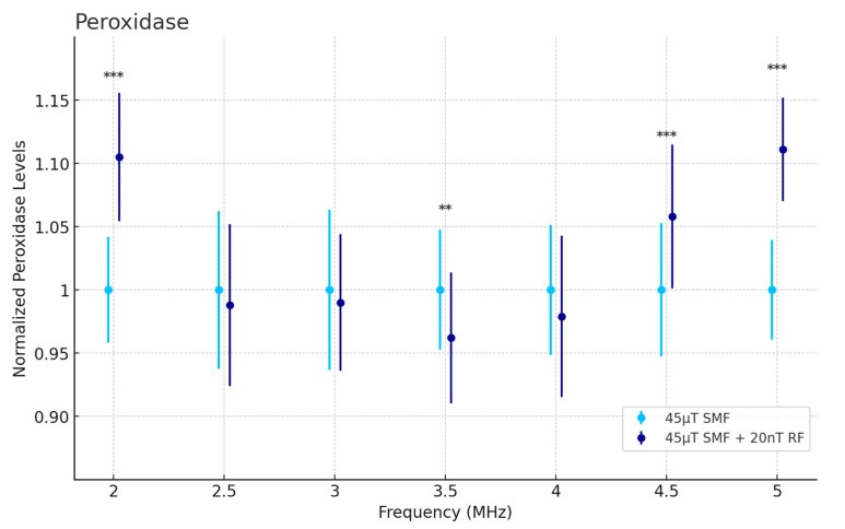
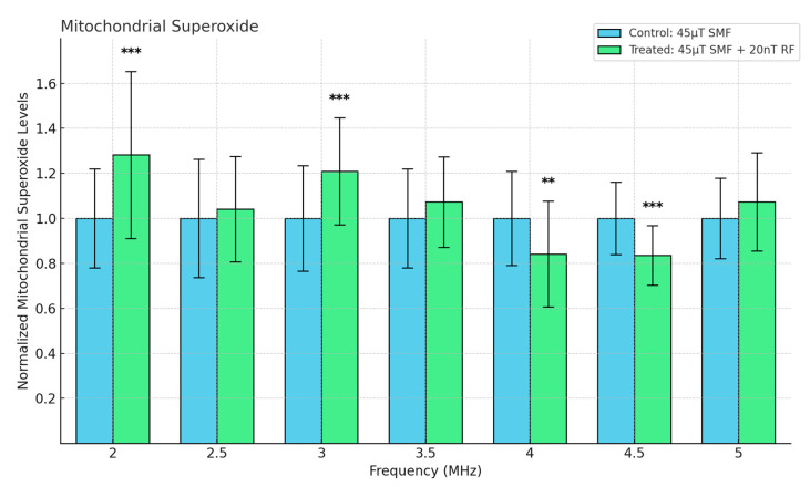
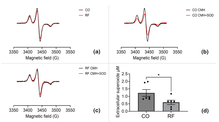
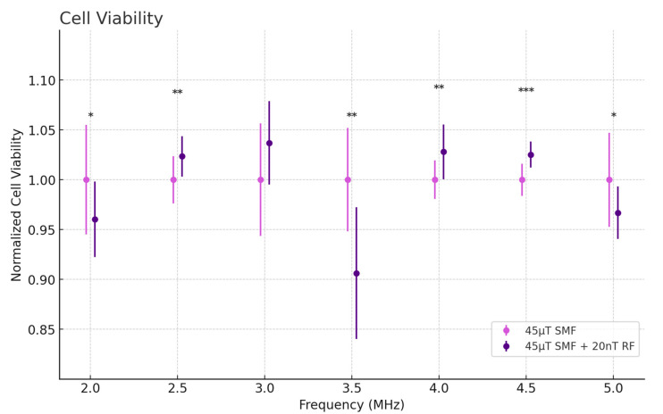
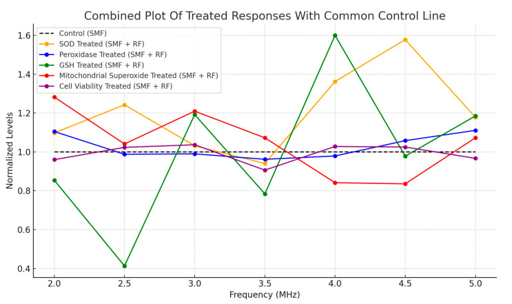
This study explores the complex relationship between radio frequency (RF) exposure and cancer cells, focusing on the HT-1080 human fibrosarcoma cell line. We investigated the modulation of reactive oxygen species (ROS) and key antioxidant enzymes, including superoxide dismutase (SOD), peroxidase, and glutathione (GSH), as well as mitochondrial superoxide levels and cell viability. Exposure to RF fields in the 2-5 MHz range at very weak intensities (20 nT) over 4 days resulted in distinct, frequency-specific cellular effects. Significant increases in SOD and GSH levels were observed at 4 and 4.5 MHz, accompanied by reduced mitochondrial superoxide levels and enhanced cell viability, suggesting improved mitochondrial function. In contrast, lower frequencies like 2.5 MHz induced oxidative stress, evidenced by GSH depletion and increased mitochondrial superoxide levels. The findings demonstrate that cancer cells exhibit frequency-specific sensitivity to RF fields even at intensities significantly below current safety standards, highlighting the need to reassess exposure limits. Additionally, our analysis of the radical pair mechanism (RPM) offers deeper insight into RF-induced cellular responses. The modulation of ROS and antioxidant enzyme activities is significant for cancer treatment and has broader implications for age-related diseases, where oxidative stress is a central factor in cellular degeneration. The findings propose that RF fields may serve as a therapeutic tool to selectively modulate oxidative stress and mitochondrial function in cancer cells, with antioxidants playing a key role in mitigating potential adverse effects.

Keywords: antioxidant enzymes; cancer cells; mitochondrial superoxide; oxidative stress; peroxidase; radical pair mechanism; radio frequency fields; reactive oxygen species; reduced glutathione; superoxide dismutase.


Conflict of interest statement

The authors declare no conflicts of interest.

Figures

Figure 1
Figure 1
Interconnected activities of antioxidant enzymes like SOD, catalase, and peroxidase, and how they mitigate oxidative stress. Adapted from [35].
Figure 2
Figure 2
X-band ESEEM spectroscopy illustrates (a) the [3Fe–4S] cluster in fumarate reductase from E. coli; (b) the [2Fe–2S] ferredoxin from A. platensis; (c) the Rieske protein in bovine heart mitochondria. Adapted from [12].
Figure 3
Figure 3
The experimental setup features two Helmholtz coils placed within a mu-metal cage. The left coil, with a single pair of turns, generates a SMF for the control cells. In contrast, the right coil consists of an inner coil that produces SMF, and an outer coil designed to generate an RF magnetic field for the treated cells. A mu-metal sheet separates the two coils within the cage, ensuring proper field isolation.
Figure 4
Figure 4
Cellular SOD levels as a function of frequency in fibrosarcoma cells. Data are expressed as mean ± SD (n = 18, N = 2) for each group. * p < 0.05, ** p < 0.01, and *** p < 0.001 represent significant differences.
Figure 5
Figure 5
Cellular GSH levels as a function of frequency in fibrosarcoma cells. Data are expressed as mean ± SD (n = 50, N = 2) for each group. *** p < 0.001 represents significant difference.
Figure 6
Figure 6
Peroxidase activity assay assessing cellular H2O2 levels as a function of frequency in fibrosarcoma cells. Data are expressed as mean ± SD (n = 36, N = 2) for each group. ** p < 0.01, and *** p < 0.001 represent significant differences.
Figure 7
Figure 7
Mitochondrial Superoxide levels as a function of frequency in fibrosarcoma cells. Data are expressed as mean ± SD (n = 32, N = 2) for each group. ** p < 0.01, and *** p < 0.001 represent significant differences.
Figure 8
Figure 8
Extracellular superoxide levels in HT-1080 cells exposed to magnetic fields. CO refers to cells exposed to a 45 µT SMF, while RF indicates cells exposed to a 45 µT SMF combined with a 20 nT RF field at 4 MHz. (a) EPR spectra of CO cells (black trace) and RF-exposed cells (red trace). (b) EPR spectra of CO cells with (red) or without (black) pretreatment with SOD1. (c) EPR spectra of RF-exposed cells, with (red) or without (black) pretreatment with SOD1. The CM● concentration was determined by double integration, followed by analysis using SpinCount. (d) SOD-inhibitable signal reflecting extracellular superoxide levels. Data are expressed as mean ± SEM; * p < 0.05 represents significant difference.
Figure 9
Figure 9
Cell Viability levels as a function of frequency in fibrosarcoma cells. Data are expressed as mean ± SD (n = 28, N = 2) for each group. * p < 0.05, ** p < 0.01, and *** p < 0.001 represent significant differences.
Figure 10
Figure 10
(a) HT-1080 cells exposed to a 45 µT SMF and 4 MHz RF field for 4 days (treated). (b) Confluence analysis of the cells shown in (a), with red contours outlining areas of low or no cell coverage. The confluency of this representative sample was calculated to be 72.77%. (c) HT-1080 cells exposed to a 45 µT SMF for 4 days (control). (d) Confluence analysis of the cells shown in (c), with red contours marking regions of low or no cell coverage. The confluency of this representative sample was calculated to be 60.05%.
Figure 11
Figure 11
Combined plot of normalized biological responses to SMF and RF exposures across 2–5 MHz. Responses include SOD, GSH, mitochondrial superoxide, and cell viability, with the control condition exposed to SMF only.

Similar articles

References

    1. Moulder J.E., Foster K.R., Erdreich L.S., McNamee J.P. Mobile Phones, Mobile Phone Base Stations and Cancer: A Review. Int. J. Radiat. Biol. 2005;81:189–203. doi: 10.1080/09553000500091097. - DOI - PubMed
    1. Habash R.W.Y., Elwood J.M., Krewski D., Lotz W.G., McNamee J.P., Prato F.S. Recent Advances in Research on Radiofrequency Fields and Health: 2004–2007. J. Toxicol. Environ. Health Part B. 2009;12:250–288. doi: 10.1080/10937400903094125. - DOI - PubMed
    1. Romanenko S., Begley R., Harvey A.R., Hool L., Wallace V.P. The Interaction between Electromagnetic Fields at Megahertz, Gigahertz and Terahertz Frequencies with Cells, Tissues and Organisms: Risks and Potential. J. R. Soc. Interface. 2017;14:20170585. doi: 10.1098/rsif.2017.0585. - DOI - PMC - PubMed
    1. Lambert N., Chen Y.-N., Cheng Y.-C., Li C.-M., Chen G.-Y., Nori F. Quantum Biology. Nat. Phys. 2013;9:10–18. doi: 10.1038/nphys2474. - DOI
    1. Arndt M., Juffmann T., Vedral V. Quantum Physics Meets Biology. HFSP J. 2009;3:386–400. doi: 10.2976/1.3244985. - DOI - PMC - PubMed

Grants and funding

LinkOut - more resources