. 2024 Oct 17;13(10):1250.
doi: 10.3390/antiox13101250.

Aged Gut Microbiome Induces Metabolic Impairment and Hallmarks of Vascular and Intestinal Aging in Young Mice

Affiliations

Aged Gut Microbiome Induces Metabolic Impairment and Hallmarks of Vascular and Intestinal Aging in Young Mice

Chak-Kwong Cheng et al. Antioxidants (Basel). .

Abstract

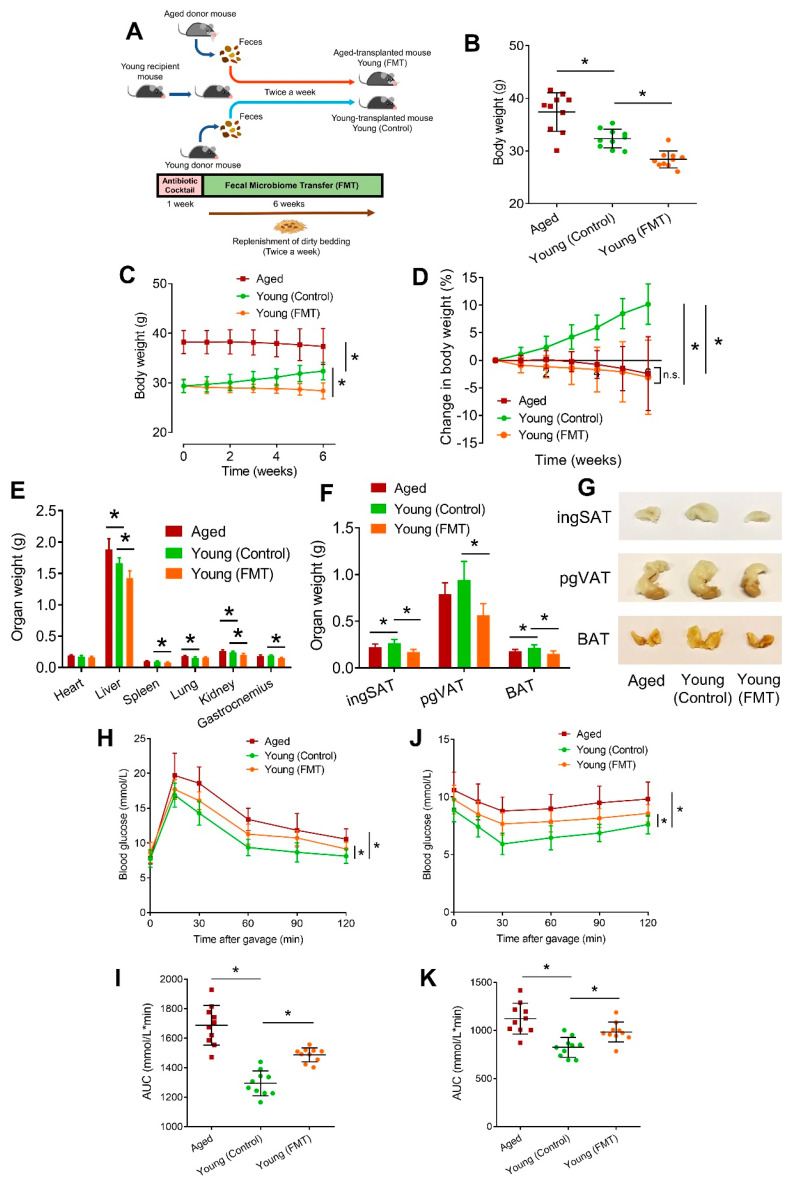
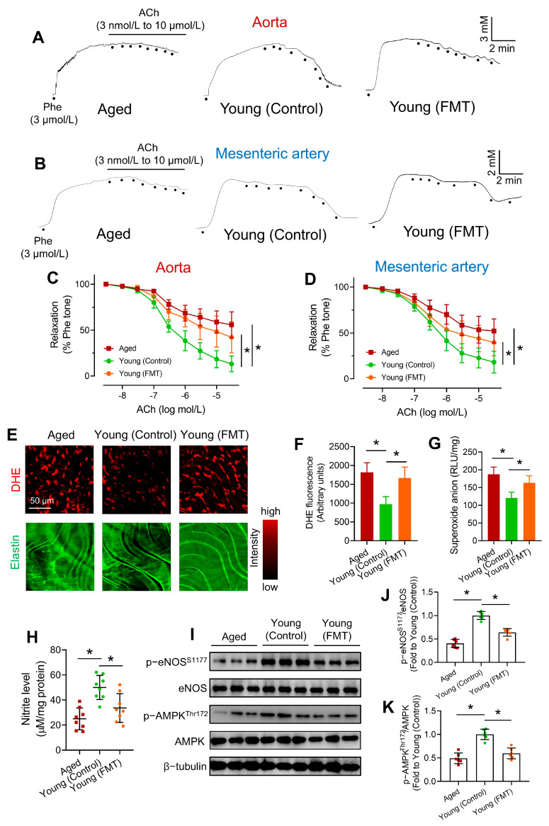
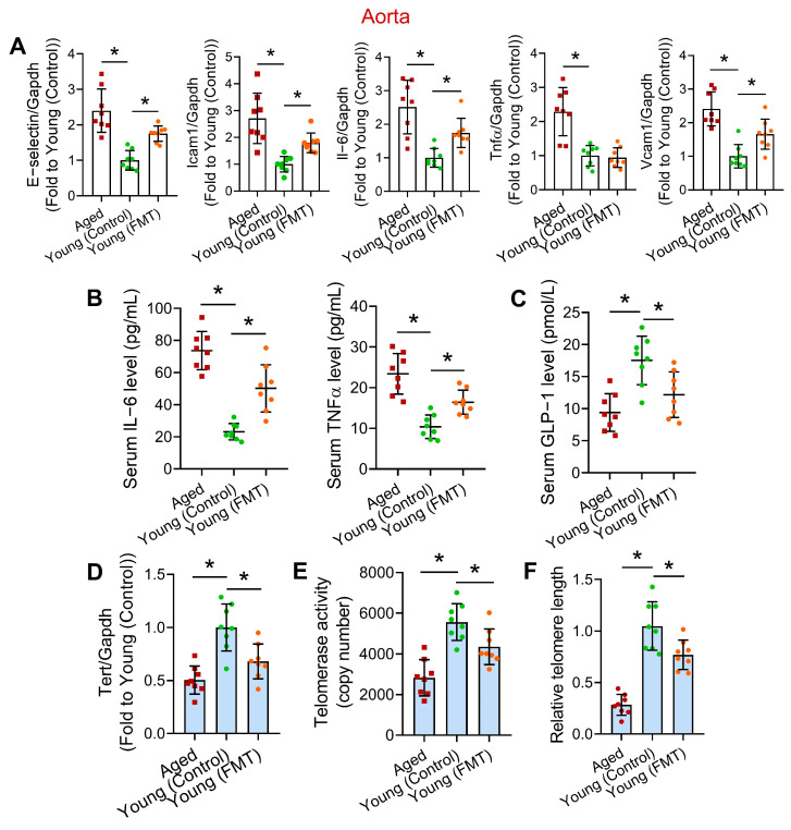
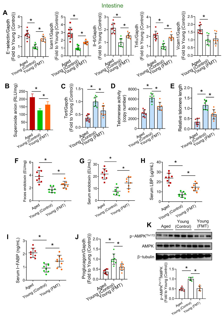
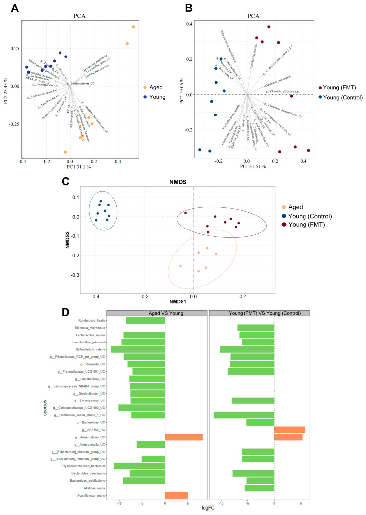
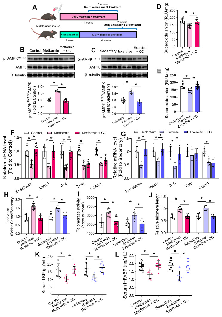
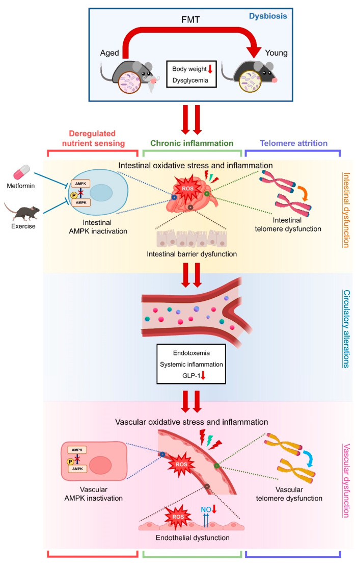
Aging, an independent risk factor for cardiometabolic diseases, refers to a progressive deterioration in physiological function, characterized by 12 established hallmarks. Vascular aging is driven by endothelial dysfunction, telomere dysfunction, oxidative stress, and vascular inflammation. This study investigated whether aged gut microbiome promotes vascular aging and metabolic impairment. Fecal microbiome transfer (FMT) was conducted from aged (>75 weeks old) to young C57BL/6 mice (8 weeks old) for 6 weeks. Wire myography was used to evaluate endothelial function in aortas and mesenteric arteries. ROS levels were measured by dihydroethidium (DHE) staining and lucigenin-enhanced chemiluminescence. Vascular and intestinal telomere function, in terms of relative telomere length, telomerase reverse transcriptase expression and telomerase activity, were measured. Systemic inflammation, endotoxemia and intestinal integrity of mice were assessed. Gut microbiome profiles were studied by 16S rRNA sequencing. Some middle-aged mice (40-42 weeks old) were subjected to chronic metformin treatment and exercise training for 4 weeks to evaluate their anti-aging benefits. Six-week FMT impaired glucose homeostasis and caused vascular dysfunction in aortas and mesenteric arteries in young mice. FMT triggered vascular inflammation and oxidative stress, along with declined telomerase activity and shorter telomere length in aortas. Additionally, FMT impaired intestinal integrity, and triggered AMPK inactivation and telomere dysfunction in intestines, potentially attributed to the altered gut microbial profiles. Metformin treatment and moderate exercise improved integrity, AMPK activation and telomere function in mouse intestines. Our data highlight aged microbiome as a mechanism that accelerates intestinal and vascular aging, suggesting the gut-vascular connection as a potential intervention target against cardiovascular aging and complications.

Keywords: AMPK; aging; dysbiosis; endothelial cell; microbiome; telomeres.


Conflict of interest statement

The authors declare no conflicts of interest.

Figures

Figure 1
Figure 1
Effects of aged-to-young FMT on body parameters. (A) Schematic overview on FMT protocol from aged and young donor mice to young recipient mice. (B) Body weights of aged donor mice (Aged), young-transplanted (Young (Control)) and aged-transplanted young mice (Young (FMT)) after 6-week FMT protocol. (C) Body weight changes and (D) percentage changes in body weights of mice in (B) during the 6-week FMT. (E) Weights of indicated organs of mice in (B) postmortem after the 6-week FMT. (F) Weights of inguinal subcutaneous adipose tissue (ingSAT), perigonadal visceral adipose tissue (pgVAT) and brown adipose tissue (BAT) of mice in (B). (G) Gross appearance of adipose tissues of mice in (B). (H) Glucose tolerance test (GTT) on mice in (B) at week 6 of FMT, and (I) corresponding area under curve (AUC) analysis of glucose over time. (J) Insulin tolerance test (ITT) of mice in (B) at week 6 of FMT, and (K) corresponding AUC analysis of glucose over time. N = 10 per group. Data are mean ± SD. * p < 0.05; Brown-Forsythe and Welch ANOVA and Dunnett T3 test.
Figure 2
Figure 2
Effects of aged-to-young FMT on endothelial function. Representative traces for endothelium-dependent relaxations (EDRs) in (A) aortas and (B) mesenteric arteries of Aged, young-transplanted (Young (Control)) and aged-transplanted mice (Young (FMT)). Summary statistics of wire myography on EDRs in (C) aortas and (D) mesenteric arteries from different mouse groups. (E) Dihydroethidium (DHE) staining on en face endothelium of different mouse groups, and (F) corresponding quantification of DHE fluorescence. (G) Lucigenin-enhanced chemiluminescence on aortic ROS levels of different mouse groups. (H) Nitrite levels in aortas of different mouse groups. N = 8 per group. (I) Representative Western blots, and (J,K) quantification of Western blotting on expression of AMPK, p-AMPK at Thr172, eNOS and p-eNOS at Ser1177 in aortas of different mouse groups. N = 6 per group. Data are mean ± SD. * p < 0.05; Brown-Forsythe and Welch ANOVA and Dunnett T3 test.
Figure 3
Figure 3
Effects of aged-to-young FMT on vascular and systemic inflammation, and vascular telomere function. (A) RT-PCR on mRNA levels of pro-inflammatory genes in aortas of Aged, young-transplanted (Young (Control)) and aged-transplanted mice (Young (FMT)). (B) ELISA on circulating inflammatory markers of different mouse groups. (C) ELISA on circulating GLP-1 levels of different mouse groups. (D) Tert mRNA level in aortas of different mouse groups. (E) Telomerase activities in aortas of different mouse groups. (F) Relative telomere length in aortas of different mouse groups. N = 8 per group. Data are mean ± SD. * p < 0.05; Brown-Forsythe and Welch ANOVA and Dunnett T3 test.
Figure 4
Figure 4
Effects of aged-to-young FMT on intestinal inflammation, telomere function and barrier function. (A) RT-PCR on mRNA levels of pro-inflammatory genes in intestines of Aged, young-transplanted (Young (Control)) and aged-transplanted mice (Young (FMT)). (B) Lucigenin-enhanced chemiluminescence on intestinal ROS levels of different mouse groups. (C) Tert mRNA level in intestines of different mouse groups. (D) Telomerase activities in intestines of different mouse groups. (E) Relative telomere length in intestines of different mouse groups. Endotoxin levels in (F) feces and (G) sera of different mouse groups. ELISA on serum levels of (H) LBP and (I) I-FABP of different mouse groups. (J) Proglucagon mRNA level in intestines of different mouse groups. N = 8 per group. (K) Representative Western blots and quantification of Western blotting on expression of AMPK and p-AMPK at Thr172 in intestines of different mouse groups. N = 6 per group. Data are mean ± SD. * p < 0.05; Brown-Forsythe and Welch ANOVA and Dunnett T3 test.
Figure 5
Figure 5
Effects of aged-to-young FMT on gut microbial profiles in young host mice. (A) Principal component analysis (PCA) plot revealing distinct clusters for fecal microbiome samples obtained from young (depicted in blue) and aged (in brown) mice before antibiotic treatment and FMT, highlighting the species contributing to this clustering. N = 8 per group. (B) PCA plot showing the clustering of fecal microbiome samples from young-transplanted (Young (Control); depicted in blue) and aged-transplanted mice (Young (FMT); in red). N = 8 per group. (C) Non-Metric Multi-Dimensional Scaling (NMDS) plot displaying the clustering of microbiome across various mouse groups. N = 6–8 per group. (D) Differential abundance analysis on the mean difference in centered log ratio for enriched species in young, aged, Young (Control) and Young (FMT) mice. N = 8 per group.
Figure 6
Figure 6
Effects of chronic metformin treatment and moderate exercise training on intestinal homeostasis. (A) Schematic diagram on chronic metformin treatment and moderate exercise training with the presence and absence of compound C (CC) treatment in middle-aged C57BL/6 mice. Representative Western blots and quantification of Western blotting on expression of AMPK and p-AMPK at Thr172 in intestines of (B) metformin-treated mice, and (C) exercise-trained mice. Lucigenin-enhanced chemiluminescence on intestinal ROS levels of (D) metformin-treated mice, and (E) exercise-trained mice. RT-PCR on mRNA levels of pro-inflammatory genes in intestines of (F) metformin-treated mice, and (G) exercise-trained mice. (H) Tert mRNA level in intestines of different mouse groups. (I) Telomerase activities in intestines of different mouse groups. (J) Relative telomere length in intestines of different mouse groups. ELISA on serum levels of (K) LBP and (L) I-FABP of different mouse groups. N = 6 per group. Data are mean ± SD. * p < 0.05; Brown-Forsythe and Welch ANOVA and Dunnett T3 test.
Figure 7
Figure 7
Schematic overview of the study. Aged microbiome induces metabolic impairments and vascular dysfunction in young mice. Aged microbiome causes telomere dysfunction, oxidative stress, and inflammation in intestines and vasculature of young mice. Metformin and moderate exercise potentially retard hallmarks of intestinal aging through AMPK activation. The study highlights the network among multiple aging hallmarks, including dysbiosis, deregulated nutrient sensing, chronic inflammation and telomere attrition, in terms of gut-vascular connection.

References

    1. Kirkwood T.B.L. Understanding the Odd Science of Aging. Cell. 2005;120:437–444. doi: 10.1016/j.cell.2005.01.027. - DOI - PubMed
    1. López-Otín C., Blasco M.A., Partridge L., Serrano M., Kroemer G. Hallmarks of Aging: An Expanding Universe. Cell. 2023;186:243–278. doi: 10.1016/j.cell.2022.11.001. - DOI - PubMed
    1. Donato A.J., Machin D.R., Lesniewski L.A. Mechanisms of Dysfunction in the Aging Vasculature and Role in Age-Related Disease. Circ. Res. 2018;123:825–848. doi: 10.1161/CIRCRESAHA.118.312563. - DOI - PMC - PubMed
    1. Ragonnaud E., Biragyn A. Gut Microbiota as the Key Controllers of “Healthy” Aging of Elderly People. Immun. Ageing. 2021;18:2. doi: 10.1186/s12979-020-00213-w. - DOI - PMC - PubMed
    1. Cheng C.K., Huang Y. The Gut-Cardiovascular Connection: New Era for Cardiovascular Therapy. Med. Rev. 2021;1:23–46. doi: 10.1515/mr-2021-0002. - DOI - PMC - PubMed

LinkOut - more resources