. 2024 Oct 18;13(10):1265.
doi: 10.3390/antiox13101265.

Rosmarinic Acid Attenuates Salmonella enteritidis-Induced Inflammation via Regulating TLR9/NF-κB Signaling Pathway and Intestinal Microbiota

Affiliations

Rosmarinic Acid Attenuates Salmonella enteritidis-Induced Inflammation via Regulating TLR9/NF-κB Signaling Pathway and Intestinal Microbiota

Dandan Yi et al. Antioxidants (Basel). .

Abstract

Salmonella enteritidis (SE) infection disrupts the homeostasis of the intestinal microbiota, causing an intestinal inflammatory response and posing a great threat to human and animal health. The unreasonable use of antibiotics has led to an increase in the prevalence of drug-resistant SE, increasing the difficulty of controlling SE. Therefore, new drug strategies and research are urgently needed to control SE. Rosmarinic acid (RA) is a natural phenolic acid with various pharmacological activities, including antioxidant, anti-inflammatory and antibacterial properties. However, the protective effects and mechanism of RA on intestinal inflammation and the gut microbial disorders caused by SE have not been fully elucidated. In this study, RAW264.7 cells, MCECs and BALB/c mice were challenged with SE to assess the protective effects and mechanisms of RA. The results showed that RA enhanced the phagocytic ability of RAW264.7 cells, reduced the invasion and adhesion ability of SE in MCECs, and inhibited SE-induced inflammation in cells. Moreover, RA inhibited the activation of the NF-κB signaling pathway by upregulating TLR9 expression. Importantly, we found that RA provided protection against SE and increased the diversity and abundance of the intestinal microbiota in mice. Compared with infection control, RA significantly increased the abundance of Firmicutes and Acidibacteria and decreased the abundance of Proteobacteria, Epsilonbacteraeota and Bacteroidota. However, RA failed to alleviate SE-induced inflammation and lost its regulatory effects on the TLR9/NF-κB signaling pathway after destroying the gut microbiota with broad-spectrum antibiotics. These results indicated that RA attenuated SE-induced inflammation by regulating the TLR9/NF-κB signaling pathway and maintaining the homeostasis of the gut microbiota. Our study provides a new strategy for preventing SE-induced intestinal inflammation.

Keywords: Gut microbiota; NF-κB; Rosmarinic acid; Salmonella enteritidis; TLR9.


Conflict of interest statement

The authors declare that they have no conflicts of interest.

Figures

Figure 1
Figure 1
RA alleviated the cellular inflammation caused by SE. (A,B) Effects of RA on the viability of RAW264.7 cells and MCECs. The cells were treated with different concentrations of RA for 6 h, 12 h, 24 h, or 48 h, and a CCK-8 assay was used to detect cell viability. The cell survival rate data at the same time were obtained for statistical analysis (n = 6). (C,D) The fluorescence images and intensity of the phagocytic ability of RAW264.7 cells. The cells were pretreated with different concentrations of RA and challenged with FITC-labeled SE (MOI = 100) for 2 h (n = 3). (E) The bacteria adhering to MCECs. The cells were pretreated with different concentrations of RA, challenged with SE (MOI = 100) for 2 h and lysed with PBS containing 0.1% Triton X-100. The bacteria adhering to the cells were detected (n = 6). (F) The intracellular bacteria in MCECs. The cells were treated with RA and challenged with SE (MOI = 100) for 2 h, and then treated with DMEM containing 1% penicillin and streptomycin for 1 h. The cells were subsequently lysed with PBS containing 1% Triton X-100 and the intracellular bacteria were detected (n = 6). (G) Levels of TNF-α, IL-1β and IL-6 in RAW264.7 cells (n = 6). (H) Levels of TNF-α, IL-1β and IL-6 in MCECs (n = 6). Cells were pretreated with different concentrations of RA for 6 h, and challenged with SE (MOI = 100) for 2 h. Cytokine levels in the cell supernatant were detected via ELISA. Statistical analysis was performed by one-way ANOVA followed by Tukey’s multiple comparison test, and expressed as the mean ± SD, * p < 0.05, ** p < 0.01, *** p < 0.001, **** p < 0.0001 versus the SE infection control group, ### p < 0.001, #### p < 0.0001 versus the blank control group.
Figure A1
Figure A1
The protective effect of RA on SE-infected mice. Organ indices of the liver, kidney and spleen (n = 6). Statistical analysis was performed by one-way ANOVA followed by Tukey’s multiple comparison test, and expressed as the mean ± SD, * p < 0.05, ** p < 0.01, *** p < 0.001, **** p < 0.0001 versus the SE infection control group.
Figure A2
Figure A2
RA regulated gut microbiota in SE-infected mice. (A): Feature numbers; (B): rare curve of the OTU level; (C): Venn diagram; (D): non-metric multi-dimensional scaling analysis; (E): linear discriminant analysis effect size.
Figure A3
Figure A3
Broad-spectrum antibiotic pretreatment significantly reduced the number of intestinal bacteria in mice. The broad-spectrum antibiotics used included neomycin (100 mg/L), streptomycin (50 mg/L), penicillin (100 mg/L), vancomycin (50 mg/L), and metronidazole (100 mg/L). The mice were orally administered with 0.5 mL of broad-spectrum antibiotics by gavage for 7 d (n = 6). After 7 d, 10 g of mouse feces was collected under sterile conditions. The feces were diluted with 1 g/mL sterile water and cultured anaerobically and aerobically on LB medium via the dilution coating plate method. The bacterial colonies on the plate were counted after 12 h at 37 °C. Student’s t-test was used for statistical analysis. **** for p < 0.0001 versus the blank control group.
Figure 2
Figure 2
RA alleviated inflammation caused by SE via regulating the TLR9/NF-κB signaling pathway. Cells were pretreated with different concentrations of RA for 6 h and challenged with SE (MOI = 100) for 2 h; cells were collected for RT-qPCR or Western blot detection. (A) Molecular docking analysis of the binding mode and affinity of RA for TLR9. (BD) The mRNA expression levels of TLR9, TNF-α and IL-6 in RAW264.7 cells (n = 6). (EG) The mRNA expression levels of TLR9, TNF-α and IL-6 in MCECs (n = 6). (HO) The protein expression levels of TLR9, p65, p-p65, IκB-α and p-IκB-α in RAW264.7 cells and MCECs (n = 3). Statistical analysis was performed by one-way ANOVA followed by Tukey’s multiple comparison test, and expressed as the mean ± SD, * p < 0.05, ** p < 0.01, *** p < 0.001, **** p < 0.0001 versus the SE infection control group.
Figure 3
Figure 3
RA inhibited the activation of NF-κB signaling pathway by upregulating TLR9. The cells were pretreated with 10 μM E6446 (TLR9 inhibitor dihydrochloride) for 2 h, and co-incubated with RA for 6 h and then challenged with SE (MOI = 100) for 2 h. The cell supernatants were collected to detect cytokines, and the cells were collected for RT–qPCR or Western blot detection. (A,B) Levels of proinflammatory cytokines in RAW264.7 cells and MCECs (n = 6). (CE) The mRNA expression levels of TLR9, TNF-α and IL-6 in RAW264.7 cells (n = 6). (FH) The mRNA expression levels of TLR9, TNF-α and IL-6 in MCECs (n = 6). (IL) The protein expression levels of TLR9, p65, p-p65, IκB-α and p-IκB-α in RAW264.7 cells (n = 3). (MP) The protein expression levels of TLR9, p65, p-p65, IκB-α and p-IκB-α in MCECs (n = 3). For (A,B), statistical analysis was performed by one-way ANOVA followed by Tukey’s multiple comparison test, and expressed as the mean ± SD, ns p > 0.05, * p < 0.05, ** p < 0.01, *** p < 0.001, **** p < 0.0001 versus the SE infection control group. For (CP), statistical analysis was performed by one-way ANOVA followed by Tukey’s multiple comparison test, and expressed as the mean ± SD, ns p > 0.05, * p < 0.05, ** p < 0.01, *** p < 0.001, **** p < 0.0001.
Figure 4
Figure 4
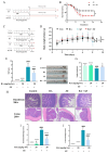
Protective effects of RA on SE-challenged mice. (A) Experimental design for evaluating the effect of RA on the survival rate of mice challenged with SE. Mice were orally pretreated with RA (20 mg/kg·bw) for 7 days and then intraperitoneally injected with 0.2 mL of SE at a concentration of 2.5 × 10⁸ CFUs/mL. The mortality of mice was recorded. (B) Survival rate of the mice (n = 20). (C) Experimental design for testing the protective effect of RA against SE. The treatment and challenge of the mice were the same as those in (A), and samples were collected 24 h after challenge. (D) Changes in the body weights of the mice (n = 10). (E) Disease activity index (n = 10). (F,G) Measurement of colon length (n = 6). (H) The histopathological changes in duodenum and colon (n = 3). (I) Duodenum histopathological score (n = 3). (J) Colon histopathological score (n = 3). For (BD), statistical analysis was performed by Mantle-Cox test and Kruskal-Wallis test, respectively, and SE infection control group was compared with other groups. In (GJ), statistical analysis was performed by one-way ANOVA followed by Tukey’s multiple comparison test, and expressed as the mean ± (SD), * p < 0.05, ** p < 0.01, *** p < 0.001, **** p < 0.0001 versus the SE infection control group, # p < 0.05, #### p < 0.0001 versus the blank control group.
Figure 5
Figure 5
RA alleviated SE-induced intestinal inflammation by regulating TLR9 and inhibiting the activation of the NF-κB signaling pathway. The mice were orally pretreated with RA (20 mg/kg·bw) for 7 days, 0.2 mL of SE at a concentration of 2.5 × 10⁸ CFUs/mL was intraperitoneally injected, and samples were collected 24 h after challenge. (AD) Serum levels of GOT, GPT, MDA and SOD (n = 6). (E) Levels of TNF-α, IL-1β and IL-6 in the serum (n = 6) (FH). The mRNA expression levels of TLR9, TNF-α and IL-6 in colon tissues (n = 6). (IL) The protein expression levels of TLR9, p65, p-p65, IκB-α and p-IκB-α in colon tissues (n = 3). Statistical analysis was performed by one-way ANOVA followed by Tukey’s multiple comparison test, and expressed as the mean ± SD, * p < 0.05, ** p < 0.01, *** p < 0.001, **** p < 0.0001 versus the SE infection control group, # p < 0.05, ## p < 0.01, #### p < 0.0001 versus the blank control group.
Figure 6
Figure 6
RA regulated the gut microbiota in SE-challenged mice (n = 6). The mice were orally pretreated with RA (20 mg/kg·bw) for 7 days, then intraperitoneally injected with 0.2 mL of SE at a concentration of 2.5 × 10⁸ CFUs/mL. (A) Circos diagram. (B) Alpha diversity analysis: observed OTUs. (C) Alpha diversity analysis: Shannon index. (D) Alpha diversity analysis: Chao 1. (E) β-diversity analysis: PCoA. (F) Relative abundance of the gut microbiota at the genus level. (GK) Relative abundance of p__Proteobacteria, p__Epsilonbacteraeota, p__Bacteroidota, p__Firmicutes and p__Actinobacteria. (L) Relative abundance of the gut microbiota at the genus level. (MR) Relative abundance of g__Muribaculaceae_unclassified, g__Bilophila, g__Tyzzerella, g__Lachnospiraceae_NK4A136_group, g__Acetatifactor and g__Intestinimonas. Statistical analysis was performed by one-way ANOVA followed by Tukey’s multiple comparison test, and expressed as the mean ± SD, * p < 0.05, ** p < 0.01, *** p < 0.001, **** p < 0.0001 versus the SE infection control group, # p < 0.05, ## p < 0.01, ### p < 0.001, #### p < 0.0001 versus the blank control group.
Figure 7
Figure 7
RA lost its protective effect against SE after clearance of the microbiota. (A) Experimental design in mice. The mice were orally pretreated with RA (20 mg/kg·bw) or antibiotics for 7 days, 0.2 mL of SE at a concentration of 2.5 × 10⁸ CFUs/mL was intraperitoneally injected, and samples were collected 24 h after challenge (n = 10). (B) Changes in body weight (n = 10). (C) Disease activity index (n = 10). (D,E) Measurement of colon length (n = 10). (F) Organ indices of the liver, kidney and spleen (n = 10). (G) Levels of TNF-α, IL-1β and IL-6 in the serum (n = 6). (H) Histopathological changes in the duodenum (n = 3). (I) Duodenal histopathological score (n = 3). For (B), statistical analysis was performed using Kruskal-Wallis test, and the SE infection control group was compared with other groups. For (CI), statistical analysis was performed by one-way ANOVA followed by Tukey’s multiple comparison test, and expressed as the mean ± SD, * p < 0.05, ** p < 0.01, **** p < 0.0001 versus the SE infection control group, # p < 0.05, ## p < 0.01, #### p < 0.0001 versus the blank control group.
Figure 8
Figure 8
RA failed to inhibit NF-κB signaling pathway by regulating TLR9 after clearance of the microbiota. The mice were orally pretreated with RA (20 mg/ kg·bw) and antibiotics for 7 days and then intraperitoneally injected with 0.2 mL of SE at a concentration of 2.5 × 10⁸ CFUs/mL. Samples were collected 24 h after challenge. (AC) The mRNA expression levels of TLR9, TNF-α and IL-6 in colon tissues (n = 6). (DG) The protein expression levels of TLR9, p65, p-p65, IκB-α and p-IκB-α in colon tissues (n = 3). Statistical analysis was performed by one-way ANOVA followed by Tukey’s multiple comparison test, and expressed as the mean ± SD, ** p < 0.01, **** p < 0.0001 versus the SE infection control group, # p < 0.05, ## p < 0.01, ### p < 0.001, #### p < 0.0001 versus the blank control group.

References

    1. LaRock D.L., Chaudhary A., Miller S.I. Salmonellae interactions with host processes. Nat. Rev. Microbiol. 2015;13:191–205. doi: 10.1038/nrmicro3420. - DOI - PMC - PubMed
    1. Karp B.E., Leeper M.M., Chen J.C., Tagg K.A., Francois Watkins L.K., Friedman C.R. Multidrug-Resistant Salmonella Serotype Anatum in Travelers and Seafood from Asia, United States. Emerg. Infect. Dis. 2020;26:1030–1033. doi: 10.3201/eid2605.190992. - DOI - PMC - PubMed
    1. Oueslati W., Rjeibi M.R., Mhadhbi M., Jbeli M., Zrelli S., Ettriqui A. Prevalence, virulence and antibiotic susceptibility of Salmonella spp. strains, isolated from beef in Greater Tunis (Tunisia) Meat Sci. 2016;119:154–159. doi: 10.1016/j.meatsci.2016.04.037. - DOI - PubMed
    1. Lin D., Yan M., Lin S., Chen S. Increasing prevalence of hydrogen sulfide negative Salmonella in retail meats. Food Microbiol. 2014;43:1–4. doi: 10.1016/j.fm.2014.04.010. - DOI - PubMed
    1. Elbediwi M., Pan H., Biswas S., Li Y., Yue M. Emerging colistin resistance in Salmonella enterica serovar Newport isolates from human infections. Emerg. Microbes Infect. 2020;9:535–538. doi: 10.1080/22221751.2020.1733439. - DOI - PMC - PubMed

LinkOut - more resources