Review
. 2024 Oct 11;25(20):10932.
doi: 10.3390/ijms252010932.

Aging Skeletal Muscles: What Are the Mechanisms of Age-Related Loss of Strength and Muscle Mass, and Can We Impede Its Development and Progression?

Affiliations
Review

Aging Skeletal Muscles: What Are the Mechanisms of Age-Related Loss of Strength and Muscle Mass, and Can We Impede Its Development and Progression?

Thomas Gustafsson et al. Int J Mol Sci. .

Abstract

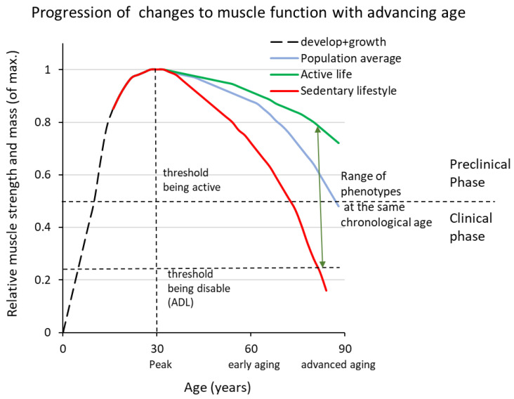
As we age, we lose muscle strength and power, a condition commonly referred to as sarcopenia (ICD-10-CM code (M62.84)). The prevalence of sarcopenia is about 5-10% of the elderly population, resulting in varying degrees of disability. In this review we emphasise that sarcopenia does not occur suddenly. It is an aging-induced deterioration that occurs over time and is only recognised as a disease when it manifests clinically in the 6th-7th decade of life. Evidence from animal studies, elite athletes and longitudinal population studies all confirms that the underlying process has been ongoing for decades once sarcopenia has manifested. We present hypotheses about the mechanism(s) underlying this process and their supporting evidence. We briefly review various proposals to impede sarcopenia, including cell therapy, reducing senescent cells and their secretome, utilising targets revealed by the skeletal muscle secretome, and muscle innervation. We conclude that although there are potential candidates and ongoing preclinical and clinical trials with drug treatments, the only evidence-based intervention today for humans is exercise. We present different exercise programmes and discuss to what extent the interindividual susceptibility to developing sarcopenia is due to our genetic predisposition or lifestyle factors.

Keywords: ageing; dynapenia; motor unit; muscle fibre atrophy; senescence.


Conflict of interest statement

The authors declare no conflict of interest.

Figures

Figure 1
Figure 1
Shows a schematic representation of the lifespan trajectory of changes in muscle strength and mass. The age at which peak performance is reached depends on the type of activity but is usually in the range of 25–35 years of age (dashed vertical line). Three curves are shown, representing very physically active individuals (green line), a person with 2–4 h of leisure-time physical activity per week (blue line) and a person with a sedentary lifestyle (red line). What all three phenotypes have in common is that peak performance is reached at around 25–30 years of age and that the rate of loss is initially slow but accelerates with advancing age. The preclinical phase continues when daily activities are not affected, while the need to moderate physical activities to cope with the loss of muscle mass and strength marks the entry into the clinical phase (the upper horizontal dashed line). As the process progresses, it eventually leads to disability (lower horizontal dashed line). Note that the range of phenotypes in the population is the product of genotype and lifestyle.
Figure 2
Figure 2
In this schematic example, a fast myofibre expressing type II myosin is denervated due to axon atrophy. In the denervated state, fast (and slow) myofibres often express both type I and type II myosins, plus embryonic myosin as a sign of denervation. When the fast myofibre is reinnervated by a slow MN, it switches to type I myosin as the predominant form, while residual amounts of type II myosin can often be detected. However, the expression of embryonic myosin is suppressed. We refer to this phenotype as hybrid myofibre. During aging, the frequency of hybrid myofibres increases from <1% to several percent of the myofibre population.
Figure 3
Figure 3
Schematic representation of the myofibre’s inherent tools that enable adaptive and regenerative responses. The interface with the tissues involved in balance and movement processes and the system level are indicated by bidirectional arrows.

References

    1. Gustafsson T., Ulfhake B. Sarcopenia: What Is the Origin of This Aging-Induced Disorder? Front. Genet. 2021;12:688526. doi: 10.3389/fgene.2021.688526. - DOI - PMC - PubMed
    1. Cai L., Shi L., Peng Z., Sun Y., Chen J. Ageing of skeletal muscle extracellular matrix and mitochondria: Finding a potential link. Ann. Med. 2023;55:2240707. doi: 10.1080/07853890.2023.2240707. - DOI - PMC - PubMed
    1. Foote K., Reinhold J., Yu E.P.K., Figg N.L., Finigan A., Murphy M.P., Bennett M.R. Restoring mitochondrial DNA copy number preserves mitochondrial function and delays vascular aging in mice. Aging Cell. 2018;17:e12773. doi: 10.1111/acel.12773. - DOI - PMC - PubMed
    1. Fleenor B.S. Large elastic artery stiffness with aging: Novel translational mechanisms and interventions. Aging Dis. 2013;4:76–83. - PMC - PubMed
    1. Plikus M.V., Wang X., Sinha S., Forte E., Thompson S.M., Herzog E.L., Driskell R.R., Rosenthal N., Biernaskie J., Horsley V. Fibroblasts: Origins, definitions, and functions in health and disease. Cell. 2021;184:3852–3872. doi: 10.1016/j.cell.2021.06.024. - DOI - PMC - PubMed

LinkOut - more resources