Review
. 2024 Oct 12;25(20):11000.
doi: 10.3390/ijms252011000.

Advances and Challenges of Bioassembly Strategies in Neurovascular In Vitro Modeling: An Overview of Current Technologies with a Focus on Three-Dimensional Bioprinting

Affiliations
Review

Advances and Challenges of Bioassembly Strategies in Neurovascular In Vitro Modeling: An Overview of Current Technologies with a Focus on Three-Dimensional Bioprinting

Salvatore Mancuso et al. Int J Mol Sci. .

Abstract

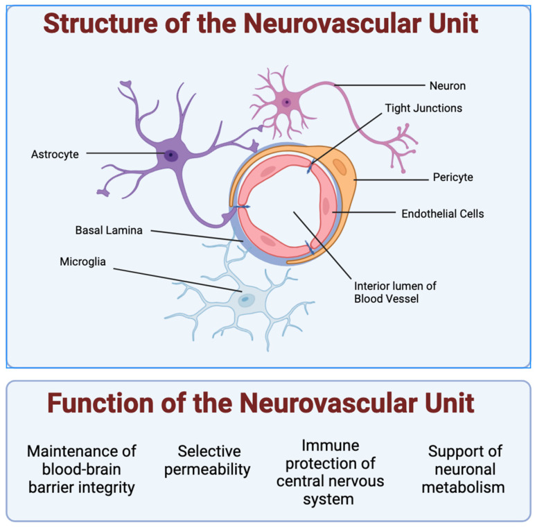
Bioassembly encompasses various techniques such as bioprinting, microfluidics, organoids, and self-assembly, enabling advances in tissue engineering and regenerative medicine. Advancements in bioassembly technologies have enabled the precise arrangement and integration of various cell types to more closely mimic the complexity functionality of the neurovascular unit (NVU) and that of other biodiverse multicellular tissue structures. In this context, bioprinting offers the ability to deposit cells in a spatially controlled manner, facilitating the construction of interconnected networks. Scaffold-based assembly strategies provide structural support and guidance cues for cell growth, enabling the formation of complex bio-constructs. Self-assembly approaches utilize the inherent properties of cells to drive the spontaneous organization and interaction of neuronal and vascular components. However, recreating the intricate microarchitecture and functional characteristics of a tissue/organ poses additional challenges. Advancements in bioassembly techniques and materials hold great promise for addressing these challenges. The further refinement of bioprinting technologies, such as improved resolution and the incorporation of multiple cell types, can enhance the accuracy and complexity of the biological constructs; however, developing bioinks that support the growth of cells, viability, and functionality while maintaining compatibility with the bioassembly process remains an unmet need in the field, and further advancements in the design of bioactive and biodegradable scaffolds will aid in controlling cell adhesion, differentiation, and vascularization within the engineered tissue. Additionally, integrating advanced imaging and analytical techniques can provide real-time monitoring and characterization of bioassembly, aiding in quality control and optimization. While challenges remain, ongoing research and technological advancements propel the field forward, paving the way for transformative developments in neurovascular research and tissue engineering. This work provides an overview of the advancements, challenges, and future perspectives in bioassembly for fabricating neurovascular constructs with an add-on focus on bioprinting technologies.

Keywords: alternatives; biomaterial; bioprinting; blood-brain barrier; cells; in vitro; matrix; neurovascular.


Conflict of interest statement

The authors declare no conflicts of interest.

Figures

Figure 1
Figure 1
Structure and function of the neurovascular unit: Shown are the 5 types of cells typically studied in the neurovascular unit. Endothelial cells form a lumen for blood to flow. Pericytes encapsulate the endothelial cells and express barrier proteins. Astrocytes and microglia monitor the junction via endfeet projections. Astrocytes help facilitate nutrient exchange to the neurons, while microglia cells act as the resident active immune defense system. The NVU supports neuronal metabolism and the healthy function of neurons in the CNS by enabling the control of oxygen and nutrient levels via vasodilation and vasoconstriction. The NVU cells also make up the blood–brain barrier (BBB), which plays a critical role in maintaining the homeostasis of the brain microenvironment as the gatekeeper of the CNS through strict and selective control of the passage of substances in and out of the brain, including the removal of waste, and protection from potentially harmful substances (endogenous and xenobiotics).
Figure 2
Figure 2
Schematic overview of current methods for organoid production. From left to right: bioreactors, hanging drop method, low-adherent plate, and the most recent, bioprinting.
Figure 3
Figure 3
An example of a microfluidic model of the neurovascular unit: Microfluidic models typically incorporate a two-chamber design; the upper channel is used for the brain partition, and the smaller, lower channel is used for the vascular partition, allowing the two distinct microenvironments to be spatially separated. A semi-permeable membrane separates the upper brain partition from the lower vessel partition. This allows for the exchanging of biomolecules and cell–cell interactions within and across the compartments. Flow induction along each channel can also be varied independently to simulate different dynamic conditions. This is only one example of the possible architectures of microfluidic chip designs. Other patterns for different use cases and cell volumes are possible.
Figure 4
Figure 4
Printing of hollow constructs using sacrificial materials: The fabrication of constructs with hollow voids is possible using a sacrificial material such as pluronic. First, the sacrificial material is printed in the desired shape of the final void. Then, the scaffold material is cast or printed atop and around the sacrificial material. Finally, the sacrificial material is removed, following the solidification of the scaffold material. This leaves a construct with a void in the negative shape of the architecture printed using the sacrificial material. This is useful for fabricating micro- and millifluidic devices and emulating larger vascular units.
Figure 5
Figure 5
Direct bioprinting: Direct bioprinting methods involve the extrusion or photo-gelation of bioink with cells directly embedded during printing. Post-processing may still be necessary.
Figure 6
Figure 6
Indirect bioprinting: Indirect bioprinting methods involve printing cell-free materials (shown in red) to support cell-laden material (shown in blue). The support material is then removed to produce the final construct.

Similar articles

Cited by

References

    1. Schlander M., Hernandez-Villafuerte K., Cheng C.Y., Mestre-Ferrandiz J., Baumann M. How Much Does It Cost to Research and Develop a New Drug? A Systematic Review and Assessment. Pharmacoeconomics. 2021;39:1243–1269. doi: 10.1007/s40273-021-01065-y. - DOI - PMC - PubMed
    1. Hackam D.G. Translating animal research into clinical benefit. BMJ. 2007;334:163–164. doi: 10.1136/bmj.39104.362951.80. - DOI - PMC - PubMed
    1. Shanks N., Greek R., Greek J. Are animal models predictive for humans? Philos. Ethics Humanit. Med. 2009;4:2. doi: 10.1186/1747-5341-4-2. - DOI - PMC - PubMed
    1. van der Worp H.B., Howells D.W., Sena E.S., Porritt M.J., Rewell S., O’Collins V., Macleod M.R. Can animal models of disease reliably inform human studies? PLoS Med. 2010;7:e1000245. doi: 10.1371/journal.pmed.1000245. - DOI - PMC - PubMed
    1. Munos B. Lessons from 60 years of pharmaceutical innovation. Nat. Rev. Drug Discov. 2009;8:959–968. doi: 10.1038/nrd2961. - DOI - PubMed

LinkOut - more resources