

Pilot Study on the Effect of Patient Condition and Clinical Parameters on Hypoxia-Induced Factor Expression: HIF1A, EPAS1 and HIF3A in Human Colostrum Cells
- PMID: 39456823
- PMCID: PMC11507067
- DOI: 10.3390/ijms252011042
Pilot Study on the Effect of Patient Condition and Clinical Parameters on Hypoxia-Induced Factor Expression: HIF1A, EPAS1 and HIF3A in Human Colostrum Cells
Abstract
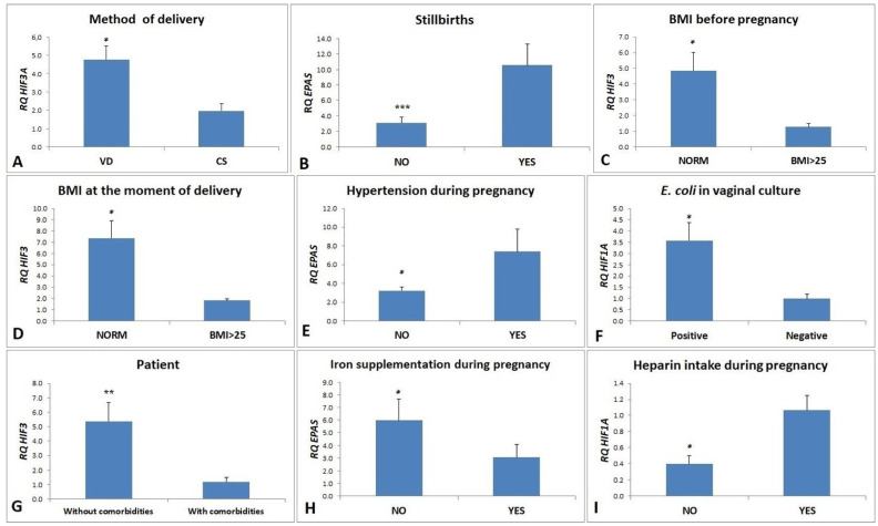
Hypoxia-inducible factor 1 (HIF-1) may play a role in mammary gland development, milk production and secretion in mammals. Due to the limited number of scientific reports on the expression of HIF genes in colostrum cells, it was decided to examine the expression of HIF1A, HIF3A and EPAS1 in the these cells, collected from 35 patients who voluntarily agreed to provide their biological material for research, were informed about the purpose of the study and signed a consent to participate in it. The expression of HIF genes was assessed using qPCR. Additionally, the influence of clinical parameters (method of delivery, occurrence of stillbirths in previous pregnancies, BMI level before pregnancy and at the moment of delivery, presence of hypertension during pregnancy, presence of Escherichia coli in vaginal culture, iron supplement and heparin intake during pregnancy) on the gene expression was assessed, revealing statistically significant correlations. The expression of HIF1A was 3.5-fold higher in the case of patients with the presence of E. coli in vaginal culture (p = 0.041) and 2.5 times higher (p = 0.031) in samples from women who used heparin during pregnancy. Approximately 1.7-fold higher expression of the EPAS1 was observed in women who did not supplement iron during pregnancy (p = 0.046). To our knowledge, these are the first studies showing the relationship between HIF expression in cells from breast milk and the method of delivery and health condition of women giving birth. The assessment of HIF expression requires deeper examination in a larger study group, and the results of further studies will allow to determine whether HIF can become biomarkers in pregnancy pathology states.
Keywords: EPAS1; HIF1A; HIF3A; colostrum; gene expression; hpoxia-inducible factor; human milk.
Conflict of interest statement
The authors declare no conflicts of interest.
Figures
Similar articles
-
HIF1A and EPAS1 potentiate hypoxia-induced upregulation of inhibin alpha chain expression in human term cytotrophoblasts in vitro.Mol Hum Reprod. 2017 Mar 1;23(3):199-209. doi: 10.1093/molehr/gax002. Mol Hum Reprod. 2017. PMID: 28115494
-
HIF1A and EPAS1 mRNA and protein expression during in vitro culture of human primary term cytotrophoblasts and effect of oxygen tension on their expression.Reprod Biol. 2016 Sep;16(3):203-211. doi: 10.1016/j.repbio.2016.05.001. Epub 2016 Jun 7. Reprod Biol. 2016. PMID: 27692362
-
miR-429 regulates the transition between Hypoxia-Inducible Factor (HIF)1A and HIF3A expression in human endothelial cells.Sci Rep. 2016 Mar 8;6:22775. doi: 10.1038/srep22775. Sci Rep. 2016. PMID: 26954587 Free PMC article.
-
Genetic variability of hypoxia-inducible factor alpha (HIFA) genes in familial erythrocytosis: Analysis of the literature and genome databases.Eur J Haematol. 2019 Oct;103(4):287-299. doi: 10.1111/ejh.13304. Epub 2019 Aug 23. Eur J Haematol. 2019. PMID: 31376207 Review.
-
EPAS1/HIF-2α is a driver of mammalian pexophagy.Autophagy. 2015;11(6):967-9. doi: 10.1080/15548627.2015.1045180. Autophagy. 2015. PMID: 25997392 Free PMC article. Review.
References
-
- Jaśkiewicz M., Moszyńska A., Króliczewski J., Cabaj A., Bartoszewska S., Charzyńska A., Gebert M., Dąbrowski M., Collawn J.F., Bartoszewski R. The transition from HIF-1 to HIF-2 during prolonged hypoxia results from reactivation of PHDs and HIF1A mRNA instability. Cell. Mol. Biol. Lett. 2022;27:109. doi: 10.1186/s11658-022-00408-7. - DOI - PMC - PubMed
MeSH terms
Substances
Grants and funding
LinkOut - more resources
Full Text Sources
