. 2024 Oct 15;25(20):11072.
doi: 10.3390/ijms252011072.

Serine Hydroxymethyltransferase 2 Deficiency in the Hematopoietic System Disrupts Erythropoiesis and Induces Anemia in Murine Models

Affiliations

Serine Hydroxymethyltransferase 2 Deficiency in the Hematopoietic System Disrupts Erythropoiesis and Induces Anemia in Murine Models

Jisheng Li et al. Int J Mol Sci. .

Abstract

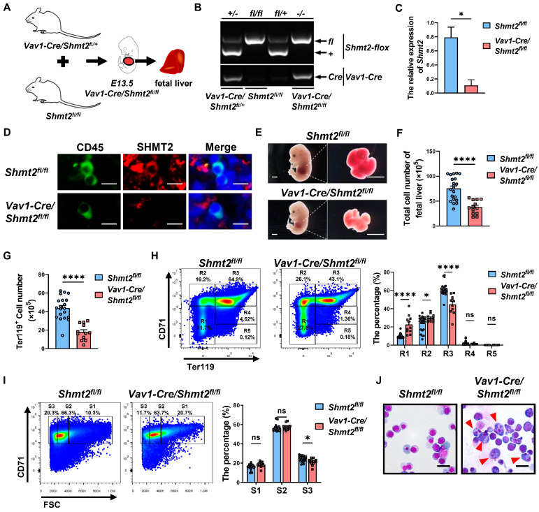
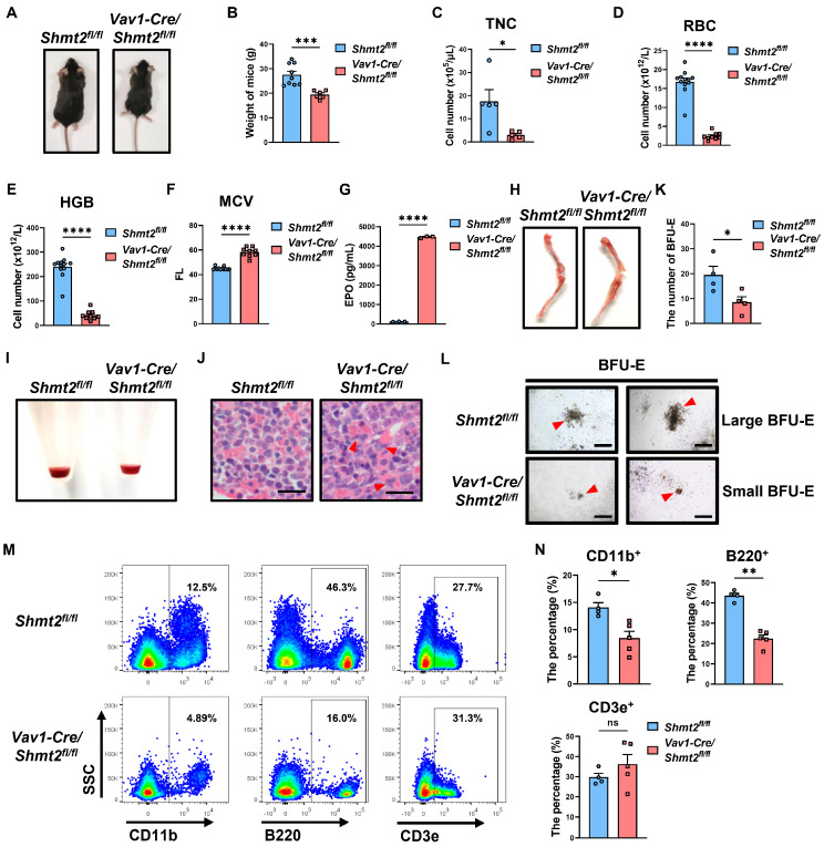
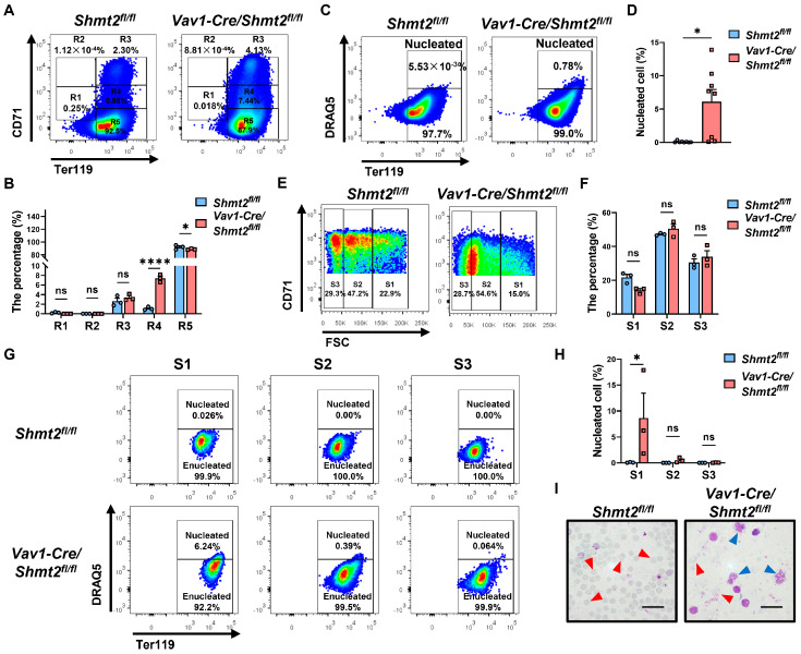
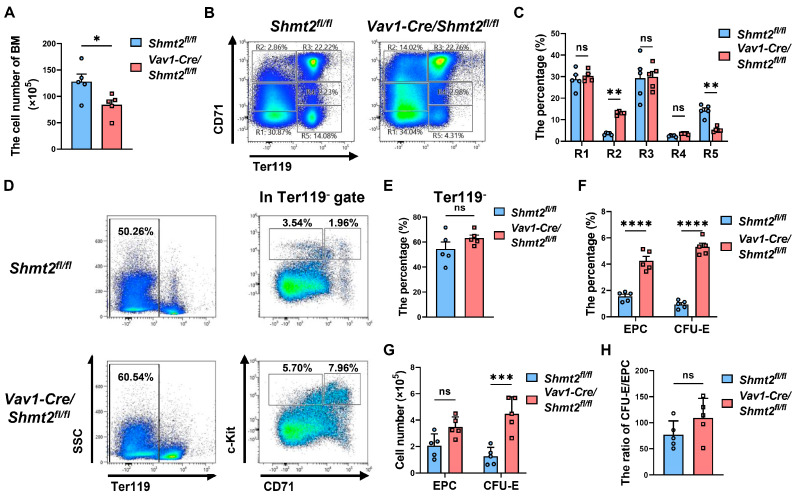
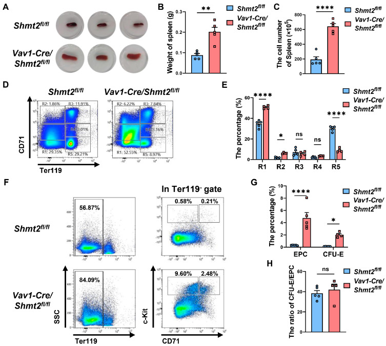
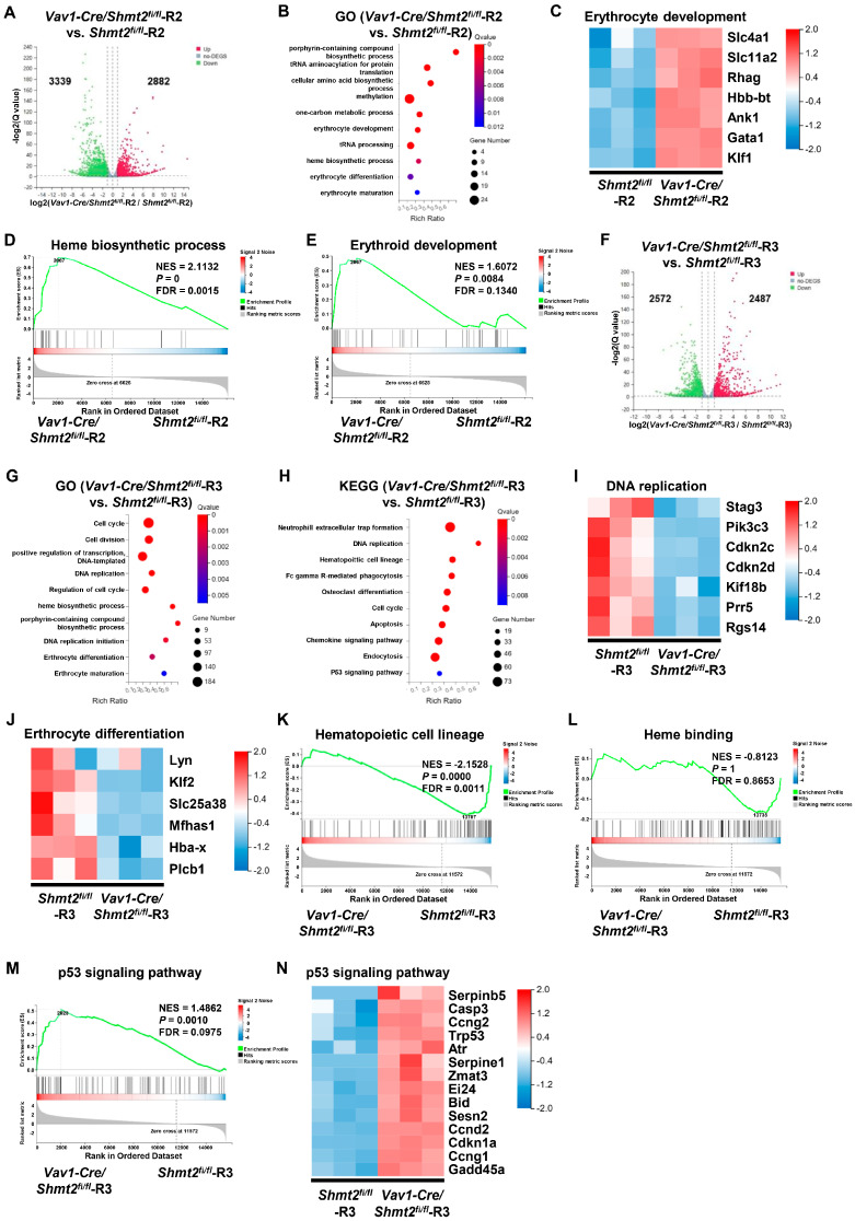
Serine and folate metabolism play critical roles in erythroid development in both embryonic and adult mice; however, the precise roles of these metabolic pathways in erythropoiesis and the pathophysiology of anemia remain inadequately characterized in the literature. To delineate the contributions of serine and folate metabolism to erythroid differentiation, we focused on serine hydroxymethyltransferase 2 (SHMT2), a key regulatory enzyme within these metabolic pathways. Using gene-editing techniques, we created fetal and adult mouse models with targeted deletion of Shmt2 in the hematopoietic system. Our findings demonstrated that the deletion of Shmt2 within the hematopoietic system led to the distinctive anemia phenotype in both fetal and adult mice. Detailed progression analysis of anemia revealed that Shmt2 deletion exerts stage-specific effects on the development and maturation of erythroid cells. Specifically, Shmt2 deficiency promoted erythroid differentiation in the R2 (CD71+ Ter119-) cell population residing in the bone marrow while concurrently inhibiting the proliferation and erythroid differentiation of the R3 (CD71+ Ter119+) cell population. This disruption resulted in developmental arrest at the R3 stage, significantly contributing to the anemia phenotype observed in the models. This study elucidates the critical role of Shmt2 in erythroid development within the hematopoietic system, highlighting the underlying mechanisms of erythroid developmental arrest associated with Shmt2 loss.

Keywords: Vav1-Cre; anemia; bone marrow; erythroid differentiation; red blood cell; serine hydroxymethyltransferase 2.


Conflict of interest statement

The authors declare no conflicts of interest.

Figures

Figure 1
Figure 1
The absence of the Shmt2 gene within the hematopoietic system impairs the production of erythroid cells in mouse fetal livers. (A). Schematic diagram of breeding Vav1-Cre/Shmt2fl/fl fetal mice with specific knockout of serine hydroxymethyltransferase 2 (Shmt2) in the hematopoietic system. (B). Agarose gel electrophoresis showing the genotyping results of Shmt2-flox (upper) and Vav1-Cre (lower) genes. (C). RT-qPCR analysis of Shmt2 expression in CD45+ cells derived from fetal livers. Statistics were determined using unpaired Student’s t-tests, * p < 0.05. n = 3. (D). Immunofluorescence staining of E13.5 fetal liver cells using anti-CD45 antibody (Alexa Fluor 488; green) and anti-SHMT2 antibody (Alexa Fluor 647; red). Scale bar: 100 μm. (E). Images of E13.5 fetal mice and fetal livers captured using a stereomicroscope. Scale bar: 1 mm. (F). Cell counting of E13.5 fetal liver cells and comparison of cell numbers between Shmt2fl/fl (n = 21) and Vav1-Cre/Shmt2fl/fl (n = 13) fetal liver cells. Statistics were determined using unpaired Student’s t-tests, **** p < 0.0001. (G). Flow cytometry analysis of the Ter119+ cell in fetal livers from Shmt2fl/fl (n = 20) and Vav1-Cre/Shmt2fl/fl (n = 12) embryos. Statistics were determined using unpaired Student’s t-tests, **** p < 0.0001. (H). Representative flow cytometry profiles of R1 to R5 erythroblast populations labeled with CD71 and Ter119 in fetal liver cells at E13.5 (left). The frequency of R1 to R5 cells in fetal livers isolated from Shmt2fl/fl (n = 25) and Vav1-Cre/Shmt2fl/fl (n = 11) embryos (right). Statistics were determined using one-way ANOVA, * p < 0.05; **** p < 0.0001; ns, not significant. (I). Representative flow cytometry profiles of Ter119hi fetal liver cells (E12.5) separated into 3 populations (S1, S2, and S3) based on the forward light scatter (FSC) profile (left). The frequency of S1 to S3 cells in fetal liver erythroid cells isolated from Shmt2fl/fl (n = 25) and Vav1-Cre/Shmt2fl/fl (n = 13) embryos (right). Statistics were determined using one-way ANOVA, * p < 0.05; ns, not significant. (J). Wright–Giemsa staining of fetal liver cells. The red arrow indicates erythroid precursor cells with irregularly shaped nuclei. Scale bar: 20 μm.
Figure 2
Figure 2
Anemia-like symptoms in adult mice with conditional Shmt2 deletion. (A). Overall view of 10-week-old Shmt2fl/fl and Vav1-Cre/Shmt2fl/fl mice. (B). Weight comparison between 10-week-old Shmt2fl/fl (n = 9) and Vav1-Cre/Shmt2fl/fl (n = 6) mice. Statistics were determined using unpaired Student’s t-tests, *** p < 0.001. (C). The total nucleated cell (TNC) counts in the peripheral blood of 10-week-old Shmt2fl/fl (n = 5) and Vav1-Cre/Shmt2fl/fl (n = 5) mice were compared. Statistics were determined using unpaired Student’s t-tests, * p < 0.05. (D). red blood cell (RBC) counts in peripheral blood of 10-week-old Shmt2fl/fl (n = 12) and Vav1-Cre/Shmt2fl/fl (n = 9) mice measured by an automatic hematology analyzer. Statistics were determined using unpaired Student’s t-tests, **** p < 0.0001. (E). hemoglobin (HGB) levels in peripheral blood of 10-week-old Shmt2fl/fl (n = 12) and Vav1-Cre/Shmt2fl/fl (n = 9) mice measured by an automatic hematology analyzer. Statistics were determined using unpaired Student’s t-tests, **** p < 0.0001. (F). mean corpuscular volume (MCV) in peripheral blood of 10-week-old Shmt2fl/fl (n = 12) and Vav1-Cre/Shmt2fl/fl (n = 9) mice measured by an automatic hematology analyzer. Statistics were determined using unpaired Student’s t-tests, **** p < 0.0001. (G). Comparison of the erythropoietin (EPO) levels in the peripheral blood serum of 10-week-old Shmt2fl/fl (n = 3) and Vav1-Cre/Shmt2fl/fl (n = 3) mice measured by ELISA. Statistics were determined using unpaired Student’s t-tests, **** p < 0.0001. (H). Images of femurs and tibias from 10-week-old Shmt2fl/fl and Vav1-Cre/Shmt2fl/fl mice. (I) Precipitates of bone marrow cells from 10-week-old Shmt2fl/fl and Vav1-Cre/Shmt2fl/fl mice. (J). H&E staining of femur sections from 10-week-old Shmt2fl/fl and Vav1-Cre/Shmt2fl/fl mice. Red arrows point to erythroid precursor cells with abnormal nuclei. Scale bar: 20 μm. (K). Quantification of burst-forming unit-erythroid (BFU-E) colonies from Methocult cultures of 1 × 10⁴ Shmt2fl/fl (n = 4) and Vav1-Cre/Shmt2fl/fl (n = 4) adult murine bone marrow cells. Statistics were determined using unpaired Student’s t-tests, * p < 0.05. Scale bar: 500 μm. (L). Large (upper) and small (lower) BFU-E colonies from Methocult cultures of 1 × 10⁴ Shmt2fl/fl and Vav1-Cre/Shmt2fl/fl adult murine bone marrow cells. Red arrows point to the BFU-E colonies. Scale bar: 500 μm. (M). Flow cytometry analysis of CD11b+, B220+, CD3e+ cell in peripheral blood (following erythrocyte lysis) from 10-week-old Shmt2fl/fl and Vav1-Cre/Shmt2fl/fl mice. (N). The frequencies of CD11b+, B220+, and CD3e+ cells in peripheral blood (following erythrocyte lysis) from 10-week-old Shmt2fl/fl (n = 4) and Vav1-Cre/Shmt2fl/fl (n = 5) mice. Statistics were determined using unpaired Student’s t-tests, * p < 0.05, ** p < 0.01, ns, not significant.
Figure 3
Figure 3
The deletion of the Shmt2 gene in the hematopoietic system impedes the enucleation of red blood cells in the peripheral blood of adult mice. (A). Representative flow cytometry profiles of R1 to R5 erythroblast populations labeled with CD71 and Ter119 in peripheral blood from 10-week-old Shmt2fl/fl and Vav1-Cre/Shmt2fl/fl mice. (B). Frequency of R1 to R5 cells in peripheral blood from 10-week-old Shmt2fl/fl (n = 3) and Vav1-Cre/Shmt2fl/fl (n = 3) mice. Statistics were determined using one-way ANOVA, * p < 0.05; **** p < 0.0001; ns, not significant. (C). Flow cytometry analysis of DRAQ5+ nucleated cells in Ter119hi peripheral blood cells from 10-week-old Shmt2fl/fl and Vav1-Cre/Shmt2fl/fl mice. (D). The frequency of DRAQ5+ nucleated cells in Ter119hi peripheral blood cells from 10-week-old Shmt2fl/fl (n = 8) and Vav1-Cre/Shmt2fl/fl (n = 8) mice. Statistics were determined using unpaired Student’s t-tests, * p < 0.05. (E). Representative flow cytograms of Ter119hi peripheral blood cells separated into 3 populations (S1, S2, and S3) based on the forward light scatter (FSC) profile. (F). The frequency of S1 to S3 cells in peripheral blood from 10-week-old Shmt2fl/fl (n = 3) and Vav1-Cre/Shmt2fl/fl (n = 3) mice. Statistics were determined using one-way ANOVA; ns, not significant. (G). Flow cytometry analysis of Ter119hi DRAQ5+ nucleated cells in peripheral blood S1 to S3 cells from 10-week-old Shmt2fl/fl and Vav1-Cre/Shmt2fl/fl mice. (H). The frequency of DRAQ5+ nucleated cells in peripheral blood S1 to S3 cells from 10-week-old Shmt2fl/fl (n = 3) and Vav1-Cre/Shmt2fl/fl (n = 3) mice. Statistics were determined using one-way ANOVA, * p < 0.05; ns, not significant. (I). Wright–Giemsa staining of peripheral blood cells from 10-week-old Shmt2fl/fl and Vav1-Cre/Shmt2fl/fl mice. Red arrows indicate enucleated erythrocytes; blue arrows indicate nucleated erythrocytes. Scale bar: 20 μm.
Figure 4
Figure 4
The absence of Shmt2 gene in the hematopoietic system hinders the development of erythroid cells in the bone marrow of adult mice. (A). BM cells counts from 10-week-old Shmt2fl/fl (n = 5) and Vav1-Cre/Shmt2fl/fl (n = 5) mice were compared. Statistics were determined using unpaired Student’s t-tests, * p < 0.05. (B). Representative flow cytometry profiles of R1 to R5 erythroblast populations labeled with CD71 and Ter119 in BM from 10-week-old adult mice. (C). The frequency of R1 to R5 cells in BM from Shmt2fl/fl (n = 5) and Vav1-Cre/Shmt2fl/fl (n = 5) 10-week-old adult mice. Statistics were determined using one-way ANOVA, ** p < 0.01; ns, not significant. (D). Representative flow cytometric results of the expression of surface markers Ter119, CD43, CD71, and c-Kit in BM cells from 10-week-old adult mice. (E,F). The bar graph shows the percentages of Ter119 cells, early progenitor cells (EPC, Ter119 CD71 c-Kit+), and colony-forming unit-erythroid (CFU-E, Ter119 CD71+ c-Kit+) cells derived from the BM cells of 10-week-old Shmt2fl/fl (n = 5) and Vav1-Cre/Shmt2fl/fl (n = 5) adult mice; Statistics were determined using unpaired Student’s t-tests, **** p < 0.0001; ns, not significant. (G). The bar graph shows the cell numbers of EPC (Ter119 CD71 c-Kit+) cells and CFU-E (Ter119 CD71+ c-Kit+) cells derived from the BM cells of 10-week-old Shmt2fl/fl (n = 5) and Vav1-Cre/Shmt2fl/fl (n = 5) adult mice. Statistics were determined using unpaired Student’s t-tests, *** p < 0.001; ns, not significant. (H). The bar graph shows the ratio of CFU-E/EPC. Statistics were determined using unpaired Student’s t-tests, ns, not significant.
Figure 5
Figure 5
The absence of the Shmt2 gene in the hematopoietic system results in splenic abnormalities in adult mice. (A). Spleen images of 10-week-old Shmt2fl/fl and Vav1-Cre/Shmt2fl/fl mice. (B). The spleens of 10-week-old Shmt2fl/fl and Vav1-Cre/Shmt2fl/fl mice were weighed, and the difference in weight between Shmt2fl/fl (n = 5) and Vav1-Cre/Shmt2fl/fl (n = 5) mice spleens was compared. Statistics were determined using unpaired Student’s t-tests, ** p < 0.01. (C). Cell counts were performed on spleen cells from 10-week-old adult mice, and the difference in spleen cell numbers between Shmt2fl/fl (n = 5) and Vav1-Cre/Shmt2fl/fl (n = 5) mice was compared. Statistics were determined using unpaired Student’s t-tests, **** p < 0.0001. (D). Representative flow cytometry profiles of R1 to R5 erythroblast populations labeled with CD71 and Ter119 in the spleens of 10-week-old adult mice. (E). The frequency of R1 to R5 cells in the spleens of Shmt2fl/fl (n = 5) and Vav1-Cre/Shmt2fl/fl (n = 5) 10-week-old adult mice. Statistics were determined using one-way ANOVA, * p < 0.05; **** p < 0.0001; ns, not significant. (F). Representative flow cytometry results of surface markers Ter119, CD43, CD71, and c-Kit in spleen cells from 10-week-old adult mice. (G). The bar graph shows the percentage of EPC (Ter119 CD71 c-Kit+) cells and CFU-E (Ter119 CD71+ c-Kit+) cells derived from the spleen cells of Shmt2fl/fl (n = 5) and Vav1-Cre/Shmt2fl/fl (n = 5) 10-week-old adult mice. Statistics were determined using unpaired Student’s t-tests, * p < 0.05; **** p < 0.0001. (H). The bar graph shows the ratio of CFU-E/EPC. Statistics were determined using unpaired Student’s t-tests, ns, not significant.
Figure 6
Figure 6
The deletion of the Shmt2 gene impedes the proliferation and erythroid differentiation of orthochromatic erythroblasts in mouse bone marrow. (A). Volcano plot showing significantly changed genes between Vav1-Cre/Shmt2fl/fl-R2 and Shmt2fl/fl-R2. Red dots represent upregulated genes. Green dots represent downregulated genes. Gray dots represent genes that were not differentially expressed (p < 0.05, |logFC| > 1.0). (B). Gene Ontology (GO) enrichment analysis revealed the top terms enriched by the upregulated genes in Vav1-Cre/Shmt2fl/fl-R2 compared to Shmt2fl/fl-R2. The size of each dot is based on the number of genes enriched in the pathway, and the color of the dots represents the significance of the pathway enrichment. (C). Heatmap illustrating the relative expression of the RNA-seq data of erythrocyte development-related genes. (D). Gene Set Enrichment Analysis (GSEA) comparing Vav1-Cre/Shmt2fl/fl-R2 and Shmt2fl/fl-R2 for heme biosynthetic process differentiation gene sets. (E). GSEA comparing Vav1-Cre/Shmt2fl/fl-R2 and Shmt2fl/fl-R2 for erythrocyte development differentiation gene sets. (F). Volcano plot showing significantly changed genes between Vav1-Cre/Shmt2fl/fl-R3 and Shmt2fl/fl-R3. Red dots represent upregulated genes. Green dots represent downregulated genes. Gray dots represent genes that were not differentially expressed (p < 0.05, |logFC| > 1.0). (G). GO enrichment analysis revealed the top terms enriched by the upregulated genes in Vav1-Cre/Shmt2fl/fl-R3 compared to Shmt2fl/fl-R3. The size of each dot is based on the number of genes enriched in the pathway, and the color of the dots represents the significance of pathway enrichment. (H). Kyoto Encyclopedia of Genes and Genomes (KEGG) enrichment analysis revealed the top terms enriched by the upregulated genes in Vav1-Cre/Shmt2fl/fl-R3 compared to Shmt2fl/fl-R3. The size of each dot is based on the number of genes enriched in the pathway, and the color of the dots represents the significance of pathway enrichment. (I). Heatmap illustrating the relative expression of the RNA-seq data of DNA replication-related genes. (J). Heatmap illustrating the relative expression of the RNA-seq data of erthrocyte differentiation-related genes. (K). GSEA comparing Vav1-Cre/Shmt2fl/fl-R3 and Shmt2fl/fl-R3 for hematopoietic cell lineage differentiation gene sets. (L). GSEA comparing Vav1-Cre/Shmt2fl/fl-R3 and Shmt2fl/fl-R3 for heme binding differentiation gene sets. (M). GSEA comparing Vav1-Cre/Shmt2fl/fl-R3 and Shmt2fl/fl-R3 for p53 signaling pathway differentiation gene sets. (N). Heatmap illustrating the relative expression of the RNA-seq data of p53 signaling pathway-related genes.

Similar articles

References

    1. Ashorobi D., Munakomi S. Myelophthisic Anemia. StatPearls Publishing; Treasure Island, FL, USA: 2023. - PubMed
    1. Stabler S.P. Clinical practice. Vitamin B12 deficiency. N. Engl. J. Med. 2013;368:149–160. doi: 10.1056/NEJMcp1113996. - DOI - PubMed
    1. Green R., Datta Mitra A. Megaloblastic anemias: Nutritional and other causes. Med. Clin. N. Am. 2017;101:297–317. doi: 10.1016/j.mcna.2016.09.013. - DOI - PubMed
    1. Green R. Vitamin B12 deficiency from the perspective of a practicing hematologist. Blood. 2017;129:2603–2611. doi: 10.1182/blood-2016-10-569186. - DOI - PubMed
    1. Socha D.S., DeSouza S.I., Flagg A., Sekeres M., Rogers H.J. Severe megaloblastic anemia: Vitamin deficiency and other causes. Clevel. Clin. J. Med. 2020;87:153–164. doi: 10.3949/ccjm.87a.19072. - DOI - PubMed

LinkOut - more resources