. 2024 Oct 17;25(20):11155.
doi: 10.3390/ijms252011155.

Eutopic and Ectopic Endometrial Interleukin-17 and Interleukin-17 Receptor Expression at the Endometrial-Myometrial Interface in Women with Adenomyosis: Possible Pathophysiology Implications

Affiliations

Eutopic and Ectopic Endometrial Interleukin-17 and Interleukin-17 Receptor Expression at the Endometrial-Myometrial Interface in Women with Adenomyosis: Possible Pathophysiology Implications

Le-Tien Hsu et al. Int J Mol Sci. .

Abstract

Adenomyosis involves the infiltration of endometrial glands and stroma deep into the uterine tissue, causing disruption to the endometrial-myometrial interface (EMI). The role of interleukin-17 (IL-17) has been extensively studied in endometriosis, but its involvement in adenomyosis remains unclear. This study aimed to investigate the expression of IL-17 in eutopic and ectopic endometrium (adenomyosis) of individuals with adenomyosis at the level of EMI. Paired tissues of eutopic endometrium and adenomyoma were collected from 16 premenopausal women undergoing hysterectomy due to adenomyosis. The IL-17 system was demonstrated in paired tissue samples at the level of EMI by the immunochemistry study. Gene expression levels of IL-17A and IL-17 receptor (IL-17R) were assessed through quantitative real-time reverse transcription polymerase chain reaction (RT-PCR). Comparative gene transcript amounts were calculated using the delta-delta Ct method. By immunohistochemical staining, CD4, IL-17A, and IL-17R proteins were detected in both eutopic endometrium and adenomyosis at the level of EMI. IL-17A and IL-17R were expressed mainly in the glandular cells, and the expression of both IL-17A and IL-17R was found to be stronger in adenomyosis than in endometrium. 3-Diaminobenzidine (DAB) staining revealed greater IL-17A expression in adenomyosis compared to eutopic endometrium. Quantitative RT-PCR showed 7.28-fold change of IL-17A and 1.99-fold change of IL-17R, and the fold change level of both IL-17A and IL-17R is significantly higher in adenomyosis (IL-17A: p = 0.047, IL-17R: p = 0.027) versus eutopic endometrium. We found significantly higher IL-17 levels in adenomyosis compared to eutopic endometrium at the level of EMI. The results showed that the IL-17 system may play a role in adenomyosis.

Keywords: adenomyosis; cytokine; endometrial–myometrial interface; interleukin-17.


Conflict of interest statement

The authors declare no conflicts of interest.

Figures

Figure 1
Figure 1
Immunohistochemistry of the IL-17 system in patients with adenomyosis. (A) Endometrial–myometrial interface stained by H&E, and the circled area indicates the adenomyotic lesion. (B,C) Negative controls without staining: (B) endometrium and (C) adenomyosis. The brown stain indicated infiltration of CD4-positive immune cells in both glandular and stromal tissues of endometrium (D) and adenomyosis (E). The brown stain (red arrow) showed the IL-17A positive reaction mainly in the glandular cells of endometrium (F) and adenomyosis (G), and the dyeing is denser in adenomyosis (G) than in endometrium (F). The brown stain (red arrow) showed the IL-17R positive reaction mainly in the glandular cells of endometrium (H) and adenomyosis (I), and the dye is denser in adenomyosis (I) than in endometrium (H). (A) scale bar = 400 μm, (B,C) scale bar = 50 μm, (DI) scale bar = 25 μm. H&E: hematoxylin and eosin.
Figure 2
Figure 2
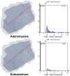
Histogram. After the paired tissues are stained by DAB, 5 areas are randomly circled at both the eutopic endometrium and adenomyosis positions. In both paired tissues, the histograms show the density information for H&E and DAB stain separately. DAB: 3,3-diaminobenzidine; H&E: hematoxylin and eosin.
Figure 3
Figure 3
Bar graph presentation of the fold change of IL-17A (A) and IL-17R (B) transcript quantified by real-time RT-PCR. Data presented are mean ± standard deviation. (A) IL-17A levels are significantly elevated in the adenomyosis group compared to the eutopic endometrium group (fold change = 7.28, p = 0.047). (B) IL-17R levels are significantly higher in the adenomyosis group compared to the eutopic endometrium group (fold change = 1.99, p = 0.027). * p < 0.05. RT-PCR: reverse transcription polymerase chain reaction.
Figure 4
Figure 4
Flow chart of study subjects.

References

    1. Upson K., Missmer S.A. Epidemiology of Adenomyosis. Semin. Reprod. Med. 2020;38:89–107. doi: 10.1055/s-0040-1718920. - DOI - PMC - PubMed
    1. Bulun S.E., Yildiz S., Adli M., Wei J.J. Adenomyosis Pathogenesis: Insights from Next-Generation Sequencing. Hum. Reprod. Update. 2021;27:1086–1097. doi: 10.1093/humupd/dmab017. - DOI - PMC - PubMed
    1. García-Solares J., Donnez J., Donnez O., Dolmans M.M. Pathogenesis of Uterine Adenomyosis: Invagination or Metaplasia? Fertil. Steril. 2018;109:371–379. doi: 10.1016/j.fertnstert.2017.12.030. - DOI - PubMed
    1. Bird C.C., McElin T.W., Manalo-Estrella P. The Elusive Adenomyosis of the Uterus--Revisited. Am. J. Obstet. Gynecol. 1972;112:583–593. doi: 10.1016/0002-9378(72)90781-8. - DOI - PubMed
    1. Zhai J., Vannuccini S., Petraglia F., Giudice L.C. Adenomyosis: Mechanisms and Pathogenesis. Semin. Reprod. Med. 2020;38:129–143. doi: 10.1055/s-0040-1716687. - DOI - PMC - PubMed

LinkOut - more resources