. 2024 Oct 17;25(20):11183.
doi: 10.3390/ijms252011183.

Application of hs-CRP in Assessment of Tissue Inflammation Following Implantation of Biodegradable Polymer in Experiment

Affiliations

Application of hs-CRP in Assessment of Tissue Inflammation Following Implantation of Biodegradable Polymer in Experiment

Igor A Eisenach et al. Int J Mol Sci. .

Abstract

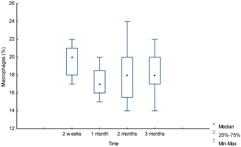
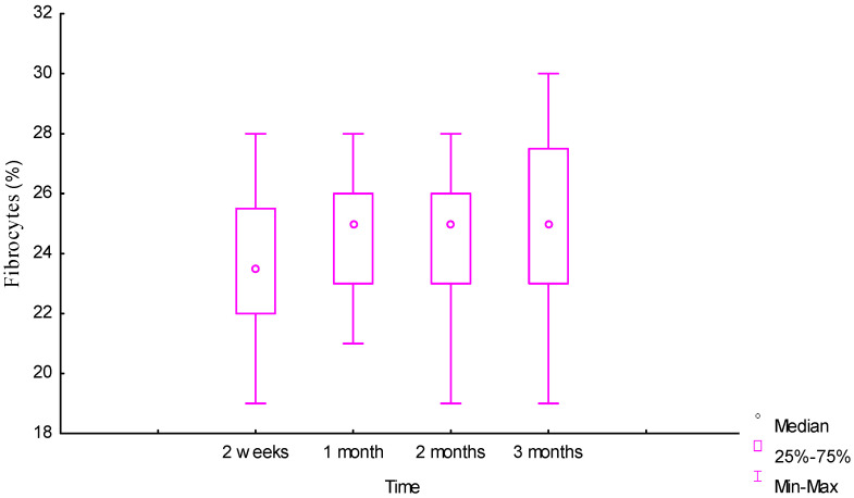
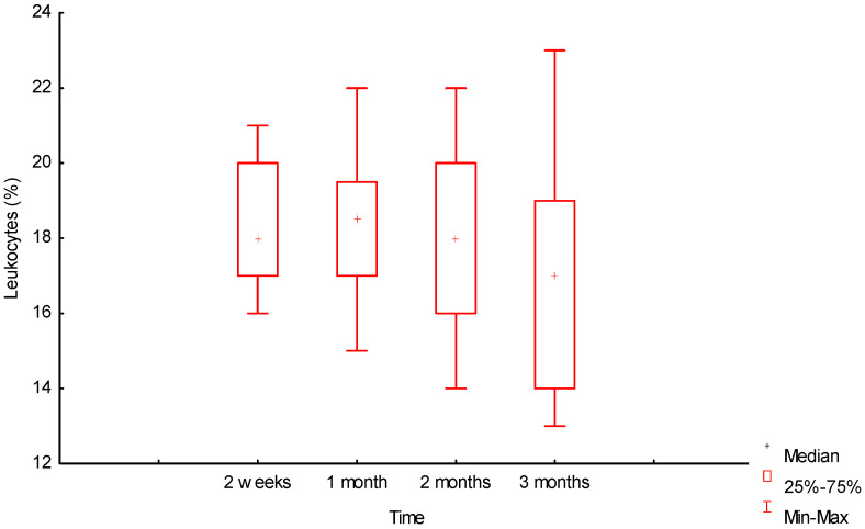
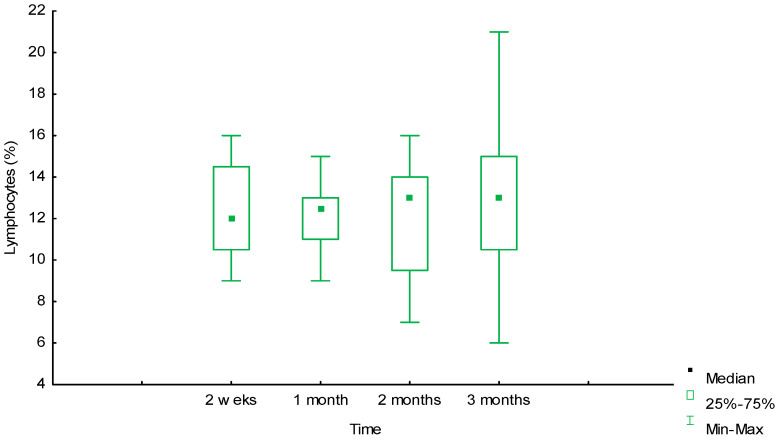
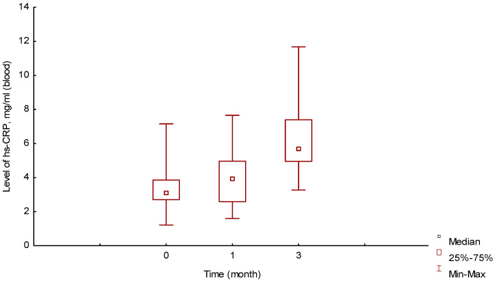
Implants made of biodegradable polymers are replaced by regenerating tissues through inflammation. The changes occurring in tissues and the organism are of practical interest for studying the biocompatibility of the material and searching for systemic markers in the blood that reflect inflammation in peri-implantation tissues. The highly sensitive C-reactive protein (hs-CRP) measurements in blood and morphometric studies of tissue surrounding the implant were carried out in the experiment within three months of implantation of a biopolymer consisting of polycaprolactone (PCL) and polytrimethylene carbonate (PTMC). During the first month, tissue inflammation decreased, and the blood level of hs-CRP did not increase. The polymer biotransformation began in the tissues after a month of implantation and was accompanied by inflammation moving deeper into the matrix. Proliferation of inflammatory cells in tissues was reflected in an increase in the hs-CRP level three months after polymer installation. The result achieved confirmed the polymer's bioinertness. The level of hs-CRP in the blood of the animals correlated with the level of inflammation in peri-implantation tissues, reflecting the activity of inflammation in the process of polymer biotransformation. This inflammation protein can be recommended for assessing tissue processes following implantation of biopolymers and their biocompatibility.

Keywords: biodegradable polymer; bioinertness; highly sensitive C-reactive protein (hs-CRP); implant; inflammation; tissue reaction.


Conflict of interest statement

The authors declare no conflicts of interest.

Figures

Figure 1
Figure 1
Biopolymer with clear boundaries in tissues, three months of implantation (macropreparation). Biopolymer matrix in the upper right corner, above the arrow.
Figure 2
Figure 2
Inflammatory infiltrate around the biopolymer, second week of the experiment. The polymer border is in the lower right corner, surrounded by an infiltrate of leukocytes, macrophages, and lymphocytes. Hematoxylin and eosin staining, magnification 100×.
Figure 3
Figure 3
Inflammatory infiltrate around the biopolymer, second month of the experiment. The polymer border is in the upper right corner (outside the picture). In the immediate vicinity of the polymer are multiple macrophages, lymphocytes, and leukocytes. Behind them are forming vessels surrounded by fibroblasts and FBGC. Hematoxylin and eosin staining, magnification 200×.
Figure 4
Figure 4
Dynamic of macrophage reaction around the biopolymer (morphometry (%) in tissues).
Figure 5
Figure 5
Dynamic of fibrocyte reaction around the biopolymer (morphometry (%) in tissues).
Figure 6
Figure 6
Dynamics of leukocyte reaction around the biopolymer (morphometry (%) in tissues).
Figure 7
Figure 7
Dynamics of lymphocyte reaction around the biopolymer (morphometry (%) in tissues).
Figure 8
Figure 8
Connective tissue around degraded biopolymer, third month of the experiment. The polymer border is in the upper right corner. Closer to it is inflammatory infiltration, behind which are many newly formed vessels, fibroblasts, and fibrocytes. In the lower left corner is dense unformed connective tissue with many collagen fibers. Van Gieson staining, magnification 200×.
Figure 9
Figure 9
Changes in blood hs-CRP levels over time, ELISA (mg/mL).
Figure 10
Figure 10
Results of biopolymer chromatography: (a) in vitro at 0 months; (b) in vitro at 2 months; (c) in vitro at 3 months; (d) in tissue, 3 months after implantation.
Figure 10
Figure 10
Results of biopolymer chromatography: (a) in vitro at 0 months; (b) in vitro at 2 months; (c) in vitro at 3 months; (d) in tissue, 3 months after implantation.

References

    1. Shtilman M.I., Artyukhov A.A., Luss A.L., Kuskov A.N. Biomaterials and regenerative medicine. Genes Cells. 2019;14:265–270.
    1. Guo B., Ma P.X. Conducting Polymers for Tissue Engineering. Biomacromolecules. 2018;19:1764–1782. doi: 10.1021/acs.biomac.8b00276. - DOI - PMC - PubMed
    1. MacCraith E., O’Brien F.J., Davis N.F. Biodegradable materials for surgical management of stress urinary incontinence: A narrative review. Eur. J. Obstet. Gynecol. Reprod. Biol. 2021;259:153–160. doi: 10.1016/j.ejogrb.2021.02.024. - DOI - PubMed
    1. Oleksy M., Dynarowicz K., Aebisher D. Advances in Biodegradable Polymers and Biomaterials for Medical Applications-A Review. Molecules. 2023;28:6213. doi: 10.3390/molecules28176213. - DOI - PMC - PubMed
    1. Szulc M., Lewandowska K. Biomaterials Based on Chitosan and Its Derivatives and Their Potential in Tissue Engineering and Other Biomedical Applications-A Review. Molecules. 2022;28:247. doi: 10.3390/molecules28010247. - DOI - PMC - PubMed

LinkOut - more resources