. 2024 Oct 19;25(20):11254.
doi: 10.3390/ijms252011254.

Encapsulation of Dexamethasone into mRNA-Lipid Nanoparticles Is a Promising Approach for the Development of Liver-Targeted Anti-Inflammatory Therapies

Affiliations

Encapsulation of Dexamethasone into mRNA-Lipid Nanoparticles Is a Promising Approach for the Development of Liver-Targeted Anti-Inflammatory Therapies

Ignacio Rivero Berti et al. Int J Mol Sci. .

Abstract

The objective of this study was to develop two lipid nanoparticle (LNP) formulations capable of efficiently expressing a reporter mRNA while co-delivering the anti-inflammatory drug dexamethasone (DX) to reduce inflammatory side effects in protein replacement therapies. Two types of LNPs were developed, in which 25% of cholesterol was replaced by DX. These LNPs contained either 1,2-distearoyl-sn-glycero-3-phosphocholine (DSPC) or 1,2-dioleoyl-sn-glycero-3-phosphoethanolamine (DOPE) as a helper lipid. The resulting LNPs exhibited high stability, homogeneity, and near-neutral Zeta potentials. SAXS experiments confirmed DX incorporation into the LNP core, with slow in vitro DX release observed over 48 h. The LNPs achieved high mRNA encapsulation efficiency (95-100%) and effectively transfected HepG2 cells, dendritic cells, and hPBMCs. While LNPs increased cytokine release (IL-1β, TNF-α, MCP-1), LNPs-DX significantly reduced cytokine levels, demonstrating enhanced anti-inflammatory properties while maintaining mRNA expression levels. In vivo biodistribution showed predominant liver localization post-intramuscular injection, regardless of the DSPC or DOPE composition. LNPs co-loaded with mRNA and DX are promising candidates for continuous protein replacement. Due to their ability to reduce treatment-related inflammation while maintaining significant mRNA expression levels, these LNPs are perfectly suited for the treatment of liver-related metabolic diseases.

Keywords: anti-inflammatory; cytokines; dendritic cells; dexamethasone; hPBMCs; in vivo biodistribution; lipid nanoparticles; mRNA delivery.


Conflict of interest statement

M.L.C. is currently an employee at BioNTech SE (Mainz, Germany); however, the contributions from M.L.C. were made prior to his employment at BioNTech. The company had no role in the design of the study; in the collection, analysis, or interpretation of data; in the writing of the manuscript; or in the decision to publish the results.

Figures

Figure 1
Figure 1
Screening of LNPs with different compositions by increasing DX concentrations into LNPs after cholesterol replacement prepared with the microfluidic system NanoAssemblr® Spark™. The effect of DX replacement on mean size (a) and PDI (b) by DLS, as well as the encapsulation efficiency (c) of the cargo molecules (EGFP mRNA and DX), were evaluated. Scheme of cholesterol replacement by structurally related analogs (DX: dexamethasone) into the LNP architecture (created with BioRender.com) (d). Composition of the different LNP formulations in mol (%) with increasing concentrations of DX (e).
Figure 2
Figure 2
Transfection efficiency of the LNP/DX formulations in HepG2 cells (a,b) and mouse dendritic cells DC 2.4 (c,d) with a total mRNA of 1 µg/mL for 24 h. Stimulation with LNP and cytokines secretion by DCs was also determined (e). Results represent the mean (n = 3) ± SD. Abbreviations: C−: untreated cells. One-way ANOVA followed by Fisher’s LSD test was used to compare between groups: p < 0.05 (*), p < 0.01 (**), p < 0.001 (***), p < 0.0001 (****), and ns (not significant; p > 0.05).
Figure 3
Figure 3
Screening of DX concentrations into LNPs with NanoAssemblr® Ignite™. Composition of the different formulation with DSPC or DOPE helper lipids (a). The mean size (b), PDI (c), and Z potential (d) of the LNPs were determined. The encapsulation efficiency of the reporter EGFP mRNA (e) and DX (f) was calculated. The results represent the mean (n = 3) ± SD. One-way ANOVA followed by Fisher’s LSD test was used to compare between groups: p < 0.05 (*), p < 0.01 (**), p < 0.001 (***), and ns (not significant; p > 0.05).
Figure 4
Figure 4
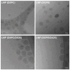
Cryo-TEM images of LNPs composed of DSPC, DOPE, DSPC/DX25, and DOPE/DX25.
Figure 5
Figure 5
SAXS profiles of the different LNP and LNP/DX formulations. The left column shows the Loglog plot of the experimental SAXS pattern in (symbol) and the fitted curve in continuous line. The right column shows the electron density profile obtained from the bilayer model in function of the radius of the particle. The density distribution is affected by the particle surface composition.
Figure 6
Figure 6
In vitro DX release from LNPs composed of DSPC and DOPE followed for 48 h. Release was performed in PBS 10 mM at pH = 7.4 and 37 °C. The kinetics were compared with the diffusion of free DX across the dialysis device. The inset shows the DX release at the first 6 h. Results are expressed as mean ± SD (n = 3).
Figure 7
Figure 7
Physical stability of DX-LNPs. LNPs demonstrated stability for at least one month (1M) when stored at 4 °C and protected from light, with no significant changes observed in size (a), PDI (b), Z pot (c), and mRNA EE% (d). Additionally, the DX-loaded LNPs showed great stability in terms of DX EE (e) and maintaining the DX release (f). The results represent the mean (n = 3) ± SD. One-way ANOVA followed by Fisher’s LSD test was used to compare among groups: ns (not significant; p > 0.05).
Figure 8
Figure 8
mRNA transfection (a) and MHC II expression (b) in dendritic cells (DC2.4) after incubation with LNPs and DX/LNPs at increasing concentrations of mRNA (1, 2, and 4 µg/mL of total mRNA) for 24 h. Mean fluorescence intensity (MFI) was analyzed by FACS. TNF-α cytokine release was also determined for all LNP formulations, suggesting that DX-loaded LNPs did not induce cytokine release, as depicted in the scheme (c). Results represent the mean (n = 3) ± SD. One-way ANOVA followed by Fisher’s LSD test was used to compare between groups: p < 0.05 (*), p < 0.01 (**), p < 0.001 (***), p < 0.0001 (****), and ns (not significant; p > 0.05).
Figure 9
Figure 9
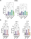
Transfection of hPBMCs with EGFP mRNA-loaded LNPs. The resulting percentage of EGFP+ cells and the mean fluorescence intensity (MFI) are shown (a). The cytokine release of TNF-α, MCP-1, and IL-1β was measured in the supernatants of stimulated hPBMCs with 8 µg/mL of mRNA loaded into LNPs (b). The results represent the mean (n = 3) ± SD. One-way ANOVA followed by Fisher’s LSD test was used to compare among groups: p < 0.05 (*), p < 0.01 (**), p < 0.001 (***), p < 0.0001 (****), and ns (not significant; p > 0.05).
Figure 10
Figure 10
Biocompatibility of DX-loaded LNPs. The cell viability of hPBMCs was determined by incorporation of the 7-AAD with flow cytometry (a). The hemotoxicity of the different LNP formulations was also assessed (b). The results represent the mean (n = 3) ± SD. One-way ANOVA followed by Fisher’s LSD test was used to compare among groups: p < 0.01 (**), p < 0.0001 (****), and ns (not significant; p > 0.05).
Figure 11
Figure 11
In vivo biodistribution of the LNPs determined in a C57BL/6 naïve mice after i.m. injection with the LNP formulations delivering Luc-mRNA. The graphs represent the mean of the region of interest (ROI) (n = 3) ± SD. One-way ANOVA followed by Fisher’s LSD test was used to compare between the groups. p < 0.05 (*), p < 0.01 (**), p < 0.0001 (****), and ns: not significant. Exposure time was 5 s.
Figure 12
Figure 12
The in vivo biodistribution at organ levels of the LNPs in C57BL/6 naïve mice after i.m. injection with the LNP formulations delivering Luc-mRNA. The graphs represent the mean (n = 3) ± SD. Two-way ANOVA test was used to compare among groups. p < 0.05 (*), p < 0.0001 (****), and ns: not significant.

References

    1. Zhang H., Han X., Alameh M.-G., Shepherd S.J., Padilla M.S., Xue L., Butowska K., Weissman D., Mitchell M.J. Rational design of anti-inflammatory lipid nanoparticles for mRNA delivery. J. Biomed. Mater. Res. A. 2022;110:1101–1108. doi: 10.1002/jbm.a.37356. - DOI - PMC - PubMed
    1. Reynolds L., Dewey C., Asfour G., Little M. Vaccine efficacy against SARS-CoV-2 for Pfizer BioNTech, Moderna, and AstraZeneca vaccines: A systematic review. Front. Public Health. 2023;11:1229716. doi: 10.3389/fpubh.2023.1229716. - DOI - PMC - PubMed
    1. Lee J., Woodruff M.C., Kim E.H., Nam J.-H. Knife’s edge: Balancing immunogenicity and reactogenicity in mRNA vaccines. Exp. Mol. Med. 2023;55:1305–1313. doi: 10.1038/s12276-023-00999-x. - DOI - PMC - PubMed
    1. Lee Y., Jeong M., Park J., Jung H., Lee H. Immunogenicity of lipid nanoparticles and its impact on the efficacy of mRNA vaccines and therapeutics. Exp. Mol. Med. 2023;55:2085–2096. doi: 10.1038/s12276-023-01086-x. - DOI - PMC - PubMed
    1. Ju Y., Carreño J.M., Simon V., Dawson K., Krammer F., Kent S.J. Impact of anti-PEG antibodies induced by SARS-CoV-2 mRNA vaccines. Nat. Rev. Immunol. 2023;23:135–136. doi: 10.1038/s41577-022-00825-x. - DOI - PMC - PubMed

LinkOut - more resources