. 2024 Oct 20;25(20):11287.
doi: 10.3390/ijms252011287.

The Therapeutic Potential of Adipose-Derived Mesenchymal Stem Cell Secretome in Osteoarthritis: A Comprehensive Study

Affiliations

The Therapeutic Potential of Adipose-Derived Mesenchymal Stem Cell Secretome in Osteoarthritis: A Comprehensive Study

Elsa González-Cubero et al. Int J Mol Sci. .

Abstract

Osteoarthritis (OA) is a degenerative joint disease characterized by cartilage degradation and inflammation. This study investigates the therapeutic potential of secretome derived from adipose tissue mesenchymal stem cells (ASCs) in mitigating inflammation and promoting cartilage repair in an in vitro model of OA. Our in vitro model comprised chondrocytes inflamed with TNF. To assess the therapeutic potential of secretome, inflamed chondrocytes were treated with it and concentrations of pro-inflammatory cytokines, metalloproteinases (MMPs) and extracellular matrix markers were measured. In addition, secretome-treated chondrocytes were subject to a microarray analysis to determine which genes were upregulated and which were downregulated. Treating TNF-inflamed chondrocytes with secretome in vitro inhibits the NF-κB pathway, thereby mediating anti-inflammatory and anti-catabolic effects. Additional protective effects of secretome on cartilage are revealed in the inhibition of hypertrophy markers such as RUNX2 and COL10A1, increased production of COL2A1 and ACAN and upregulation of SOX9. These findings suggest that ASC-derived secretome can effectively reduce inflammation, promote cartilage repair, and maintain chondrocyte phenotype. This study highlights the potential of ASC-derived secretome as a novel, non-cell-based therapeutic approach for OA, offering a promising alternative to current treatments by targeting inflammation and cartilage repair mechanisms.

Keywords: conditioned medium; inflammatory cytokines; mesenchymal stem cells; osteoarthritis; secretome.


Conflict of interest statement

The authors declare no conflict of interest.

Figures

Figure 1
Figure 1
TNF-induced modulation of inflammatory cytokines with and without CM treatment. qPCR was used to measure the relative gene expression of NOS2, IL6, MMP13 and TNF genes in non-inflamed chondrocytes, ASCs, TNF-inflamed chondrocytes and CM-treated TNF-inflamed chondrocytes after 12 h of incubation. The results are expressed as the mean ± SD of three independent experiments. * (p ≤ 0.05), ** (p ≤ 0.01) compared to non-stimulated cells ^ (p ≤ 0.05), ^^ (p ≤ 0.01) compared to TNF-stimulated cells.
Figure 2
Figure 2
Functional annotation analysis of DEGs that were significantly downregulated or upregulated (A) comparing gene expression in non-inflamed chondrocytes and TNF-stimulated chondrocytes and (B) comparing gene expression in TNF-inflamed chondrocytes and CM-treated TNF-inflamed chondrocytes.
Figure 3
Figure 3
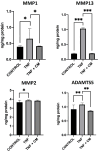
Modulation of MMP1, MMP2, MMP13, and ADAMTS5 proteins in chondrocytes. ELISA was used to measure protein levels comparing non-inflamed, TNF-inflamed, and CM-treated TNF-inflamed chondrocytes after 12 h of incubation. The results are expressed as the mean ± SD of three independent experiments. * p ≤ 0.05, ** p ≤ 0.01, *** p ≤ 0.001.
Figure 4
Figure 4
Inflammation-induced upregulation of hypertrophic factors in chondrocytes. RUNX2 and COL10A1 gene expression was determined for non-inflamed, TNF-inflamed, and CM-treated TNF-inflamed chondrocytes after 12 h of incubation (A) qPCR analysis with bone as control. The results are expressed as the mean ± SD of three independent experiments. *** p ≤ 0.001. (B) Western blot analysis with ACTB used as endogenous control. Levels of COL10A1 and RUNX2 were detected only in TNF-inflamed chondrocytes not treated with CM. (C) Immunocytochemical analysis (representative image): Blue fluorescence indicates cell nuclei (DAPI); green immunofluorescence shows presence of RUNX2 (Alexa-488); and red immunofluorescence indicates the presence of COL10A1 (Alexa-568). Scale bar: 20–50 µm. (D) Histogram of RUNX2 and COL10A1 immunofluorescence. The results are expressed as the mean ± SD of three independent experiments. *** p ≤ 0.001.
Figure 5
Figure 5
Secretome induced regeneration in inflamed chondrocytes. Gene expression for a range of chondrogenic genes was compared for non-inflamed, TNF-inflamed, and CM-treated TNF-inflamed chondrocytes after 12 h of incubation. (A) qPCR analysis of COL2A1, ACAN and SOX9. The results are expressed as the mean ± SD of three independent experiments. * p ≤ 0.05, *** p ≤ 0.001. (B) Western blot analysis of SOX9 and COL2A1; ACTB was used as endogenous control. (C) Immunohistochemical analysis of COL2A1, ACAN and SOX9 (representative image). Blue fluorescence indicates cell nuclei (DAPI); green immunofluorescence shows presence of COL2A1 (Alexa-488); red immunofluorescence indicates the presence of ACAN (Alexa-568); and green immunofluorescence shows presence of SOX9 (Alexa-488). (D) Histogram showing results for COL2A1, ACAN and SOX9 immunofluorescence. The results are expressed as the mean ± SD of three independent experiments. ** p ≤ 0.01, *** p ≤ 0.001.
Figure 6
Figure 6
Secretome induced proliferation and blocked NF-κB translocation in inflamed chondrocytes. Non-inflamed, TNF-inflamed, and CM-treated TNF-inflamed chondrocytes were compared after 12 h of incubation to assess cell proliferation and NF-κB activation in the various experimental conditions. (A) Cell proliferation (representative images). Blue fluorescence indicates cell nuclei (DAPI). Scale bar: 50 µm. (B) Cell proliferation fluorescence histograms. The results are expressed as the mean ± SD of three independent experiments. ** p ≤ 0.01. (C) NF-κB activation (representative image): Blue fluorescence indicates cell nuclei (DAPI) and green immunofluorescence indicates the presence of NF-κB p65 antibodies so enabling the localization of NF-κB p65. Scale bar: 10 µm. (D) Relative intensity of green fluorescence observed cell nuclei. The results are expressed as the mean ± SD of three independent experiments. ** p ≤ 0.01 ANOVA was used comparing TNF treated samples with all other groups.

Similar articles

Cited by

References

    1. Cui A., Li H., Wang D., Zhong J., Chen Y., Lu H. Global, Regional Prevalence, Incidence and Risk Factors of Knee Osteoarthritis in Population-Based Studies. eClinicalMedicine. 2020;29–30:100587. doi: 10.1016/j.eclinm.2020.100587. - DOI - PMC - PubMed
    1. Funck-Brentano T., Cohen-Solal M. Subchondral Bone and Osteoarthritis. Curr. Opin. Rheumatol. 2015;27:420–426. doi: 10.1097/BOR.0000000000000181. - DOI - PubMed
    1. Fernandes J.C., Martel-Pelletier J., Pelletier J.-P. The Role of Cytokines in Osteoarthritis Pathophysiology. Biorheology. 2002;39:237–246. - PubMed
    1. Tetlow L.C., Adlam D.J., Woolley D.E. Matrix Metalloproteinase and Proinflammatory Cytokine Production by Chondrocytes of Human Osteoarthritic Cartilage: Associations with Degenerative Changes. Arthritis Rheum. 2001;44:585–594. doi: 10.1002/1529-0131(200103)44:3<585::AID-ANR107>3.0.CO;2-C. - DOI - PubMed
    1. Zhang Q., Ji Q., Wang X., Kang L., Fu Y., Yin Y., Li Z., Liu Y., Xu X., Wang Y. SOX9 Is a Regulator of ADAMTSs-Induced Cartilage Degeneration at the Early Stage of Human Osteoarthritis. Osteoarthr. Cartil. 2015;23:2259–2268. doi: 10.1016/j.joca.2015.06.014. - DOI - PubMed

MeSH terms