. 2024 Oct 20;25(20):11292.
doi: 10.3390/ijms252011292.

The Expression of Cytokines and Chemokines Potentially Distinguishes Mild and Severe Psoriatic Non-Lesional and Resolved Skin from Healthy Skin and Indicates Different Stages of Inflammation

Affiliations

The Expression of Cytokines and Chemokines Potentially Distinguishes Mild and Severe Psoriatic Non-Lesional and Resolved Skin from Healthy Skin and Indicates Different Stages of Inflammation

Renáta Bozó et al. Int J Mol Sci. .

Abstract

In the psoriatic non-lesional (PS-NL) skin, the tissue environment potentially influences the development and recurrence of lesions. Therefore, we aimed to investigate mechanisms involved in regulating tissue organization in PS-NL skin. Cytokine, chemokine, protease, and protease inhibitor levels were compared between PS-NL skin of patients with mild and severe symptoms and healthy skin. By comparing mild and severe PS-NL vs. healthy skin, differentially expressed cytokines and chemokines suggested alterations in hemostasis-related processes, while protease inhibitors showed no psoriasis severity-related changes. Comparing severe and mild PS-NL skin revealed disease severity-related changes in the expression of proteases, cytokines, and chemokines primarily involving methyl-CpG binding protein 2 (MECP2) and extracellular matrix organization-related mechanisms. Cytokine and chemokine expression in clinically resolved versus healthy skin showed slight interleukin activity, differing from patterns in mild and severe PS-NL skin. Immunofluorescence analysis revealed the severity-dependent nuclear expression pattern of MECP2 and decreased expression of 5-methylcytosine and 5-hydroxymethylcytosine in the PS-NL vs. healthy skin, and in resolved vs. healthy skin. Our results suggest distinct cytokine-chemokine signaling between the resolved and PS-NL skin of untreated patients with varying severities. These results highlight an altered inflammatory response, epigenetic regulation, and tissue organization in different types of PS-NL skin with possibly distinct, severity-dependent para-inflammatory states.

Keywords: chemokines; cytokines; non-lesional skin; psoriasis; psoriasis severity; resolved skin.


Conflict of interest statement

The authors declare no competing interests.

Figures

Figure 1
Figure 1
Differential expressions of cytokines/chemokines, proteases, and protease inhibitors between healthy and non-lesional skin of psoriatic patients with mild and severe disease. The levels of cytokines/chemokines (A), proteases (B), and protease inhibitors (C) were compared in whole skin punch biopsies from healthy donors and from non-lesional skin of untreated psoriatic patients. For mild psoriasis, the mean Psoriasis Area and Severity Index (PASI) was 9.06, while for severe psoriasis, the mean PASI was 28.93. Each group utilized n = 3 pooled protein extracts. Pixel densities of the different proteins were determined by the Image Studio software 6.0 (LI-COR Biosciences, Lincoln, NE, USA) and presented as relative protein expression. In the visualization, the steel blue color corresponds to the lowest expression, red indicates the highest expression, and white represents medium expression levels of the given protein. Proteins are indicated by gene identifiers and listed in Appendix A Table A1.
Figure 2
Figure 2
Differentially expressed proteins (DEPs) and the biological pathways influenced by DEPs in both mild and severe psoriatic non-lesional skin versus healthy skin. Cytokines/chemokines, proteases, and protease inhibitors showing at least twofold changes (increase or decrease) in both mild and severe psoriatic non-lesional (PS-NL) vs. healthy (A), mild PS-NL-only vs. healthy (B), and severe PS-NL-only vs. healthy skin (C). For mild psoriasis, the mean Psoriasis Area and Severity Index (PASI) was 9.06, while for severe psoriasis, the mean PASI was 28.93. Each group utilized n = 3 pooled protein extracts. Pixel densities of the different proteins were determined by the Image Studio software (LI-COR Biosciences, Lincoln, NE, USA) and presented as relative protein expression. In the visualization, the steel blue color corresponds to the lowest expression, red indicates the highest expression, and white represents medium expression levels of the given protein. Proteins are indicated by gene identifiers and listed in Appendix A Table A1. Green squares label the cytokine/chemokine group, yellow squares indicate proteases, and grey squares mark protease inhibitors. The most significant biological pathways affected by DEPs in both mild and severe PS-NL vs. healthy skin (D), in mild PS-NL-only vs. healthy skin (E), and in severe PS-NL-only vs. healthy skin (F) were identified by the Reactome over-representation test. Results were filtered by statistical significance (p ≤ 0.05), and numbering indicates the order of significance. FDR: false discovery rate. Known data for the expression of DEPs in psoriatic lesional and non-lesional skin and their severity-related expression (G). The red arrow indicates increased expression, and the blue arrow marks decreased expression. + means known protein expression; – means that no data were found [19,20,21,22,23,24,25,26,27,28,29,30,31,32,33,34,35,36,37,38,39,40]. Comparison of the foldchange direction of DEP array data and microarray data of healthy versus non-lesional psoriatic skin (H). Matched foldchange direction is presented. PS-NL: psoriatic non-lesional.
Figure 3
Figure 3
Differentially expressed proteins (DEPs) and the biological pathways affected by DEPs in severe versus mild psoriatic non-lesional skin. DEPs with decreased (A) and increased (B) expression in the severe psoriatic non-lesional (PS-NL) vs. mild PS-NL skin are presented. Light blue dotted columns indicate mild PS-NL and blue dotted columns mark severe PS-NL pixel densities. For mild psoriasis, the mean Psoriasis Area and Severity Index (PASI) was 9.06, while for severe psoriasis, the mean PASI was 28.93. Each group utilized n = 3 pooled protein extracts. Proteins are indicated by gene identifiers and listed in Appendix A Table A1. The most relevant biological pathways affected by decreased (C) and increased (D) DEPs in the severe vs. mild PS-NL skin were analyzed by the Reactome over-representation test. Results were filtered by statistical significance (p ≤ 0.05), and numbering indicates the order of significance. FDR: false discovery rate.
Figure 4
Figure 4
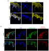
Psoriasis-severity-related reduced expression of brain-derived neurotrophic factor (BDNF), 5-methylcytosine (5mC), and 5-hydroxymethylcytosine (5hmC), and altered expression pattern of methyl-CpG binding protein 2 (MECP2) in psoriatic non-lesional versus healthy skin. Immunofluorescence staining for BDNF (a), MECP2 (b), as well as 5mC and 5hmC (c) in mild and severe psoriatic non-lesional (PS-NL) skin, and healthy skin. Mild psoriatic patients’ Psoriasis Area and Severity Index (PASI) scores were between 5.9 and 11.5; severe psoriatic patients’ PASI scores were between 20.6 and 28.93. Representative images are shown; n = 3 donors were used in each group; 40× (oil) original magnification, scale bar: 20 μm; DAPI: 4′,6-diamidino-2-phenylindole; Zeiss AxioImager Z1 microscope (Carl Zeiss AG, Oberkochen, Germany).
Figure 5
Figure 5
Distinct expression of cytokines and chemokines in clinically resolved psoriatic skin. The levels of cytokines and chemokines were compared in whole skin punch biopsies from healthy donors and resolved psoriatic skin of treated patients (n = 3 pooled protein extracts for each group) (A). The Image Studio software (LI-COR Biosciences, Lincoln, NE, USA) was used to determine the pixel densities of the different proteins. In the visualization, the blue color corresponds to the lowest expression, red indicates the highest expression, and white represents medium expression levels for the given protein. Differentially expressed cytokines and chemokines in resolved vs. healthy skin are presented by pixel densities (B). The black color indicates the healthy group, and the white, dotted pattern marks the resolved group. The most relevant biological pathways affected by these differentially expressed cytokines and chemokines in the resolved vs. healthy skin were analyzed by the Reactome over-representation test (C). DEPs of cytokines and chemokines in the mild and severe psoriatic non-lesional (PS-NL) vs. healthy skin and resolved vs. healthy skin were also compared (D). Unlabeled proteins showed decreased expression, and red-marked proteins showed increased expression. Protein expression labeled with an asterisk decreased in the mild and severe PS-NL vs. healthy skin and increased in the resolved vs. healthy skin. The Reactome test was performed to reveal the most relevant pathways of differentially expressed cytokines and chemokines 1. in both mild and severe PS-NL vs. healthy skin (E); 2. in mild PS-NL-only vs. healthy skin (F); 3. in severe PS-NL-only vs. healthy skin (G); 4. in resolved-only vs. healthy skin (H); and 5. in resolved vs. healthy skin for the *—labeled opposite expression pattern showing differentially expressed cytokines and chemokines (I). The Reactome results were filtered by statistical significance (p ≤ 0.05), and numbering indicates the order of significance. FDR: false discovery rate. Proteins are indicated by gene identifiers and listed in Appendix A Table A1. PS-NL: psoriatic non-lesional.
Figure 6
Figure 6
The expression of known psoriasis-related cytokines and chemokines in the mild and severe psoriatic non-lesional versus healthy skin is mainly reduced or non-altered but slightly increased in resolved versus healthy skin. Healthy normalized, relative pixel densities of known psoriasis-related cytokines and chemokines are presented in the mild and severe psoriatic non-lesional (PS-NL) versus healthy skin (a) and in resolved psoriatic versus healthy skin (b) using our cytokine/chemokine protein array data (n = 3 pooled protein extracts for each group). Black color indicates healthy skin, light-blue dotted columns indicate mild PS-NL, blue dotted columns mark severe PS-NL, and white dotted pattern marks the resolved group pixel densities.
Figure 7
Figure 7
Increased nuclear expression of methyl-CpG binding protein 2 (MECP2) and decreased staining patterns of 5-methylcytosine (5mC) and 5-hydroxymethylcytosine (5hmC) in resolved versus healthy skin. Immunolocalization of MECP2 (a) and 5mC and 5hmC (b) in psoriatic resolved versus healthy skin. Representative images are shown; n = 3 donors were used for each group; 40× (oil) original magnification, scale bar: 20 μm; DAPI: 4′,6-diamidino-2-phenylindole; Zeiss AxioImager Z1 microscope (Carl Zeiss AG, Oberkochen, Germany).
Figure 8
Figure 8
Different stages of inflammation in different healthy-appearing non-lesional skin types. Schematic representation of the identified processes and their potential anti-inflammatory and pro-inflammatory roles. In healthy skin, anti- and pro-inflammatory processes are in a balanced state, providing a homeostatic environment. However, in mild and severe psoriatic non-lesional and resolved psoriatic skin, where tissue stress is present, anti- and pro-inflammatory pathways may alter the balanced homeostatic state, creating a different inflammatory environment with potential distinct, severity-dependent para-inflammatory states. Yellow color indicates potential anti-inflammatory processes, and blue color marks possible pro-inflammatory processes. The illustration was created with BioRender.com.

Similar articles

Cited by

References

    1. Guo J., Zhang H., Lin W., Lu L., Su J., Chen X. Signaling pathways and targeted therapies for psoriasis. Signal Transduct. Target. Ther. 2023;8:437. doi: 10.1038/s41392-023-01655-6. - DOI - PMC - PubMed
    1. Icen M., Crowson C.S., McEvoy M.T., Dann F.J., Gabriel S.E., Maradit Kremers H. Trends in incidence of adult-onset psoriasis over three decades: A population-based study. J. Am. Acad. Dermatol. 2009;60:394–401. doi: 10.1016/j.jaad.2008.10.062. - DOI - PMC - PubMed
    1. Griffiths C.E.M., Armstrong A.W., Gudjonsson J.E., Barker J.N.W.N. Psoriasis. Lancet Lond. Engl. 2021;397:1301–1315. doi: 10.1016/S0140-6736(20)32549-6. - DOI - PubMed
    1. Nosbaum A., Dahel K., Goujon C., Nicolas J.-F., Mengeaud V., Vocanson M. Psoriasis is a disease of the entire skin: Non-lesional skin displays a prepsoriasis phenotype. Eur. J. Dermatol. EJD. 2021;31:143–154. doi: 10.1684/ejd.2021.4015. - DOI - PubMed
    1. Ghaffarinia A., Ayaydin F., Póliska S., Manczinger M., Bolla B.S., Flink L.B., Balogh F., Veréb Z., Bozó R., Szabó K., et al. Psoriatic Resolved Skin Epidermal Keratinocytes Retain Disease-Residual Transcriptomic and Epigenomic Profiles. Int. J. Mol. Sci. 2023;24:4556. doi: 10.3390/ijms24054556. - DOI - PMC - PubMed

LinkOut - more resources