. 2024 Oct 21;25(20):11305.
doi: 10.3390/ijms252011305.

α4 Nicotinic Acetylcholine Receptors in Lipopolysaccharide-Related Lung Inflammation

Affiliations

α4 Nicotinic Acetylcholine Receptors in Lipopolysaccharide-Related Lung Inflammation

Jeffrey D Ritzenthaler et al. Int J Mol Sci. .

Abstract

Sepsis remains an important healthcare challenge. The lungs are often affected in sepsis, resulting in acute lung injury characterized by inflammation. Mechanisms involving lipopolysaccharide (LPS) stimulation of toll-like receptor (TLR) signaling with induction of proinflammatory pathways have been implicated in this process. To date, however, studies targeting these pathways have failed to improve outcomes. We have found that LPS may also promote lung injury through the activation of α4 nicotinic acetylcholine receptors (α4 nAChRs) in immune cells. We observed increased expression of α4 nAChRs in human THP-1 monocytic cells exposed to LPS (100 ng/mL, 24 h). We also observed that LPS stimulated the expression of other relevant genes, including tumor necrosis factor-α, interleukin-1β, plasminogen activator inhibitor-1, the solute carrier family 7 member 11, extracellular superoxide dismutase, and transforming growth factor-β1. Of interest, dihydro-β-erythroidine hydrobromide (DHβE), a specific chemical inhibitor of α4 nAChRs, inhibited the LPS-induced expression of these genes. We generated mice with a global knockout mutation of the α4 nAChR subunit in the C57BL/6 background using CRISPR/Cas9 technology. The lungs of these LPS-treated animals demonstrated a reduction in the expression of the above-mentioned genes when compared with the lungs of wild-type animals. In support of the role of oxidative stress, we observed that LPS induced expression of the cystine transporter Slc7a11 in both THP-1 cells and in wild-type mouse lungs. The effects of LPS on THP-1 cells were blocked by the thiol antioxidant N-acetylcysteine and mimicked by redox stress. Importantly, the induction of IL-1β by redox stress was inhibited by the α4 nAChR inhibitor DHβE. Finally, we showed that LPS stimulated calcium influx in THP-1 cells, which was blocked by the α4 nAChR inhibitor. Our observations suggest that LPS promotes lung injury by stimulating redox stress, which activates α4 nAChR signaling and drives proinflammatory cytokine expression.

Keywords: lung injury; monocytic cells; nicotinic receptors; redox stress; sepsis.


Conflict of interest statement

The authors declare no conflict of interest.

Figures
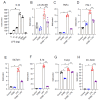
Figure 1
Figure 1
Effects of LPS in THP-1 cells. (A) Human monocytic cells, THP-1, were initially treated with varying doses of LPS (0–10,000 ng, 24 h) and found to have optimal response at 100 ng. Thus, further experiments were performed with that dose. (BH) Treatment with LPS (100 ng/mL, 24 h) showed increased mRNA expression of α4 nAChRs and several oxidative stress and inflammatory genes including TNFα, PAI-1, Slc7a11, IL-1β, TGFβ, and EC-SOD compared with non-LPS-treated control cells. Results of 2-Way ANOVA followed by post hoc analysis of comparisons between groups are shown (Tukey’s multiple comparison test). Results expressed as the fold increase in mRNA compared with samples from non-treated control cells using the 2−ΔΔCt method. * p < 0.0002 when compared with non-treated control cells. ** p < 0.0002 when compared with LPS-treated cells.
Figure 2
Figure 2
Effects of LPS in the lungs of α4 nAChR KO mice. (A) Lung tissue from LPS-treated animals (3 μg/g IT, 24 h) showed increased inflammation as demonstrated by an influx in monocytic cells (20× magnification). Some cells are depicted with arrows. (B) The findings did not differ between WT and knockout animals. (CG) Lung tissue isolated from LPS (10 mg/kg, 4 h)-treated wild-type animals showed increased expression of α4 nAChR, TNFα, PAI-1, Slc7a11, and EC-SOD mRNA when compared with control non-treated mice. The α4 nAChR KO mice treated with LPS demonstrated a significant reduction in the expression of TNFα, PAI-1, EC-SOD, and Scl7a11 when compared with wild-type animals. Data were analyzed as described for Figure 1. * p < 0.0002 when compared with non-treated control cells. ** p < 0.0002 when compared with LPS-treated cells.
Figure 3
Figure 3
THP-1 cells after LPS stimulation and exposure to redox stress. (A,B) Lung tissue isolated from LPS-treated wild-type animals showed increased expression of Slc7a11 and fibronectin EDA protein (green color; red color for control gene) when compared with untreated animals, while α4 nAChR knockout animals failed to show this effect. (C) THP-1 cells treated with LPS (100 ng/mL, 24 h) showed induction of Slc7a11 mRNA. This effect was inhibited by N-acetylcysteine (NAC, 5 mM). * p < 0.0001 when compared with non-treated control cells. ** p < 0.0001 when compared with LPS. (D) THP-1 cells treated with LPS showed induction of IL-1β mRNA expression. This effect was inhibited by N-acetylcysteine (NAC, 5 mM). * p < 0.0001 when compared with non-treated control cells. ** p < 0.0001 when compared with LPS. (E) Redox stress (Eh Cys/CySS 0 mV)-induced IL-1β, which was inhibited by the α4 nAChR inhibitor, DHβE (100 μM). * p < 0.0001 when compared with −80 mV media. ** p < 0.0001 when compared with 0 mV media.
Figure 4
Figure 4
LPS induced calcium influx in THP-1 cells. THP-1 cells were treated with LPS (100 ng/mL, 0–6 min) followed by calcium influx detection. LPS induced calcium influx within 1 min after exposure while DHβE alone had no effect. However, DHβE (100 μM) inhibited the induction of calcium influx in cells exposed to LPS. * p < 0.0002 when compared with non-treated control, DHβE, and LPS + DHβE cells.
Figure 5
Figure 5
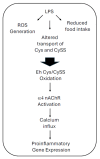
LPS-induced redox stress and α4 nAChR-dependent lung inflammation. It has been postulated that LPS induces oxidation of the plasma Cys/CySS redox state in animals by a combination of three mechanisms: early generation of reactive oxygen species, altered transport of Cys and CySS, and reduced food intake [11,12]. The resulting oxidized Eh Cys/CySS triggers α4 nAChR activation and signaling through calcium influx. The latter results in the expression of proinflammatory genes.

References

    1. Mayr F.B., Yende S., Angus D.C. Epidemiology of severe sepsis. Virulence. 2014;5:4–11. doi: 10.4161/viru.27372. - DOI - PMC - PubMed
    1. Singer M., Deitschmann C.S., Seymour C.W., Shankar-Hari M., Annane D., Bauer M., Bellomo R., Bernand G.R., Chiche J.-D., Coopersmith C.M., et al. The Third International Consensus Definitions for Sepsis and Septic Shock. JAMA. 2016;315:801–810. doi: 10.1001/jama.2016.0287. - DOI - PMC - PubMed
    1. Reinhart K., Daniels R., Kissoon N., Machado F.R., Schachter R.D., Finfer S. Recognizing sepsis as a global health priority—A WHO Resolution. N. Engl. J. Med. 2017;377:414–417. doi: 10.1056/NEJMp1707170. - DOI - PubMed
    1. Kim W.-Y., Hong S.-B. Sepsis and acute respiratory distress syndrome: Recent update. Tuberc. Respir. Dis. 2016;79:53–57. doi: 10.4046/trd.2016.79.2.53. - DOI - PMC - PubMed
    1. Domscheit H., Hegeman M.A., Carvalho N., Spieth P.M. Molecular dynamics of lipopysaccharide-induced lung injury in rodents. Front. Physiol. 2020;11:36. doi: 10.3389/fphys.2020.00036. - DOI - PMC - PubMed

MeSH terms

Substances

Grants and funding

LinkOut - more resources