. 2024 Oct 21;25(20):11307.
doi: 10.3390/ijms252011307.

Proliferative Diabetic Retinopathy Microenvironment Drives Microglial Polarization and Promotes Angiogenesis and Fibrosis via Cyclooxygenase-2/Prostaglandin E2 Signaling

Affiliations

Proliferative Diabetic Retinopathy Microenvironment Drives Microglial Polarization and Promotes Angiogenesis and Fibrosis via Cyclooxygenase-2/Prostaglandin E2 Signaling

Shuta Kishishita et al. Int J Mol Sci. .

Abstract

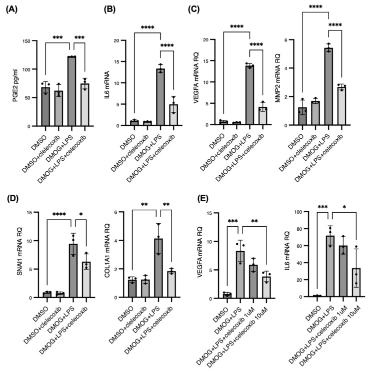
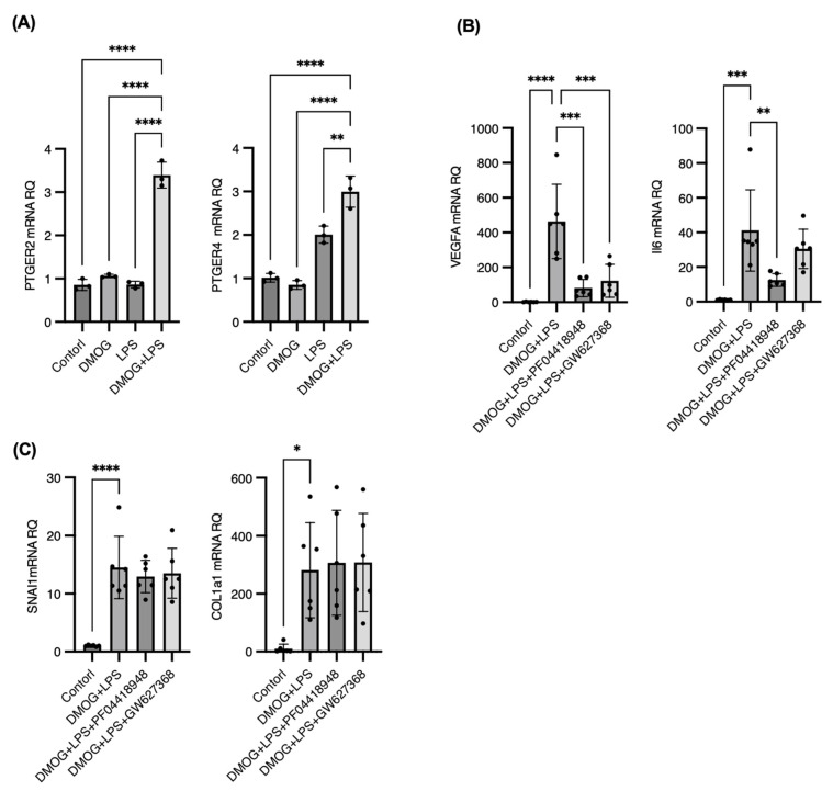
Diabetic retinopathy (DR) is the leading cause of visual impairment, particularly in the proliferative form (proliferative DR [PDR]). The impact of the PDR microenvironment on microglia, which are the resident immune cells in the central nervous system, and the specific pathological changes it may induce remain unclear. This study aimed to investigate the role of microglia in the progression of PDR under hypoxic and inflammatory conditions. We performed a comprehensive gene expression analysis using human-induced pluripotent stem cell-derived microglia under different stimuli (dimethyloxalylglycine (DMOG), lipopolysaccharide (LPS), and DMOG + LPS) to mimic the hypoxic inflammatory environment characteristic of PDR. Principal component analysis revealed distinct gene expression profiles, with 76 genes synergistically upregulated under combined stimulation. Notably, prostaglandin-endoperoxide synthase 2 (encoding cyclooxygenase (COX)-2) exhibited the most pronounced increase, leading to elevated prostaglandin E2 (PGE2) levels and driving pathological angiogenesis and inflammation via the COX-2/PGE2/PGE receptor 2 signaling axis. Additionally, the upregulation of the fibrogenic genes snail family transcriptional repressor 1 and collagen type I alpha 1 chain suggested a role for microglia in fibrosis. These findings underscore the critical involvement of microglia in PDR and suggest that targeting both the angiogenic and fibrotic pathways may present new therapeutic strategies for managing this condition.

Keywords: COX-2; diabetic retinopathy; human microglia; hypoxia; inflammation; neurovascular unit.


Conflict of interest statement

The authors declare no conflicts of interest.

Figures

Figure 1
Figure 1
Distinct global gene expression profiles in induced pluripotent stem cell-derived microglia (iMGs) under hypoxia and immune activation. Comprehensive gene expression profiles were analyzed to assess the effects of hypoxia and innate immune activation on human iMGs using lipopolysaccharide (LPS) and dimethyloxaloylglycine (DMOG) (A). The heatmap shows that DMOG stimulation resulted in the significant upregulation of 216 genes, whereas LPS stimulation upregulated 912 genes compared with the control. In addition, these differences were confirmed, highlighting a subset of genes that was synergistically upregulated by both stimuli (B). Pathway analysis of the differentially expressed genes indicated the activation of the vascular endothelial growth factor and hypoxia-inducible factor-1 signaling pathways under DMOG stimulation, indicating a hypoxic response (C). Conversely, LPS stimulation enhanced Toll-like receptor signaling, nuclear factor-kappaB signaling, and tumor necrosis factor signaling, suggesting heightened immune activation (D).
Figure 2
Figure 2
Synergistic upregulation of prostaglandin-endoperoxide synthase 2 (PTGS2) and pathological gene networks in induced pluripotent stem cell-derived microglia under hypoxia and immune activation. We found 76 genes that were significantly and synergistically upregulated by both innate immune-activating stimulation by lipopolysaccharide and hypoxic stimulation by dimethyloxaloylglycine compared with either stimulus alone (false discovery rate < 0.1, fold change > 1.5) (A). The top 20 synergistically upregulated genes are highlighted; the most significantly upregulated gene is PTGS2, which encodes cyclooxygenase 2 (B). Gene ontology analysis of the synergistically upregulated genes identified significant terms related to angiogenesis, cell migration, chemotaxis, and fibrosis (C).
Figure 3
Figure 3
Synergistic upregulation of cyclooxygenase 2 and prostaglandin E2 in response to hypoxia and inflammation. Quantitative polymerase chain reaction for prostaglandin-endoperoxide synthase 2 (PTGS2) and enzyme-linked immunosorbent assay (ELISA) for prostaglandin E2 (PGE2) and prostaglandin F2α (PGF2α) were performed under 200 μM dimethyloxaloylglycine (DMOG), 100 ng/mL lipopolysaccharide (LPS), or combined DMOG and LPS stimulation for 24 h. The expression of PTGS2 mRNA was significantly upregulated, showing a 10–20-fold increase with single LPS or DMOG stimulation and a 150–200-fold increase with combined DMOG and LPS stimulation compared with the control (A). ELISA measurements indicated increased levels of PGE2, but not PGF2α, in the culture supernatant under these conditions (B,C). Aqueous humor samples were collected from five patients with proliferative diabetic retinopathy (PDR) accompanied with diabetic macular edema (two males and three females; average age, 64.4 ± 6.2 years) and five age-matched patients without any retinal diseases as control (two males and three females; average age, 64.0 ± 10.1 years) for ELISA assay. Vitreous humor samples were also collected from five patients with PDR (four males and one female; average age, 57.6 ± 5.9 years) and five age-matched patients without any retinal diseases as control (three males and two females; average age, 61.0 ± 5.2 years). PGE2 concentrations were significantly elevated in the aqueous and vitreous humors of patients with PDR compared with those in the controls (D,E). * p < 0.05, ** p < 0.01, *** p < 0.001, **** p < 0.0001.
Figure 4
Figure 4
Synergistic activation of angiogenesis- and fibrosis-related genes in induced pluripotent stem cell-derived microglia under hypoxia and inflammation. Quantitative polymerase chain reaction for genes associated with angiogenesis and fibrovascular proliferation in proliferative diabetic retinopathy (PDR) were performed under 200 μM dimethyloxaloylglycine (DMOG), 100 ng/mL lipopolysaccharide (LPS), or combined DMOG and LPS stimulation for 24 h. The expression levels of vascular endothelial growth factor A, matrix metalloproteinase 2, and interleukin-6, key contributors to pathological neovascularization in PDR, were significantly upregulated under combined DMOG and LPS stimulation compared with monostimulation (AC). Snail family transcriptional repressor 1 (SNAI1), a master transcription factor that regulates mesenchymal transition, was upregulated under both DMOG and LPS stimulation (D). Collagen type I alpha 1 chain, a major extracellular matrix component induced by SNAI1 activation, markedly increased under combined stimulation (E). ** p < 0.01, *** p < 0.001, **** p < 0.0001.
Figure 5
Figure 5
Celecoxib inhibits cyclooxygenase 2 (COX-2) mediated inflammatory, angiogenic, and fibrotic gene expression in induced pluripotent stem cell-derived microglia (iMGs) under hypoxia and inflammation. We treated iMGs stimulated by 200 μM dimethyloxaloylglycine (DMOG) and 100 ng/mL lipopolysaccharide (LPS) with 10 μM celecoxib, the selective COX-2 inhibitor, for 24 h. Celecoxib treatment significantly downregulated prostaglandin E2 production in the culture supernatant (A). The quantitative polymerase chain reaction results showed that celecoxib markedly reduced the expression of interleukin-6 (IL-6) in both DMOG and LPS-stimulated iMGs (B). Celecoxib significantly decreased the expression of vascular endothelial growth factor A (VEGFA) and matrix metalloproteinase 2, the critical drivers of pathological neovascularization, which were notably upregulated under combined DMOG and LPS stimulation (C). The expression of snail family transcriptional repressor 1, a key transcription factor for fibrosis, and its downstream target, collagen type I alpha 1 chain, was significantly reduced by celecoxib treatment (D). The expression of VEGFA and IL-6, which was elevated by DMOG + LPS, was suppressed in a dose-dependent manner by celecoxib (E). * p < 0.05, ** p < 0.01, *** p < 0.001, **** p < 0.0001.
Figure 6
Figure 6
The cyclooxygenase 2/prostaglandin E2/PGE receptor 2 (EP2) axis as a central driver of angiogenesis and inflammation in induced pluripotent stem cell-derived microglia (iMGs), with distinct pathways governing fibrosis. The genes encoding prostaglandin receptors EP2 (PTGER2) and EP4 (PTGER4) were expressed in human iMGs and were notably upregulated by 200 μM dimethyloxaloylglycine (DMOG) and 100 ng/mL lipopolysaccharide (LPS) stimulation for 24 h, whereas the genes encoding EP1 (PTGER1) and EP3 (PTGER3) were not expressed (A). To pinpoint which prostaglandin receptors drive the upregulation of angiogenic and fibrogenic genes, we treated iMGs with specific antagonists PF04418948, targeting EP2, and GW627368, targeting EP4 receptors, under combined 200 μM DMOG and 100 ng/mL LPS stimulation for 24 h. Treatment of iMGs with PF04418948 significantly downregulated vascular endothelial growth factor A and interleukin-6 mRNA expression, underscoring the pivotal role of the EP2 receptor in mediating these pathways (B). However, SNAI1 and COL1A1 did not respond to EP2 or EP4 antagonists, indicating that they have different regulatory mechanisms (C). * p < 0.05, ** p < 0.01, *** p < 0.001, **** p < 0.0001.

Similar articles

Cited by

References

    1. Wykoff C.C., Khurana R.N., Nguyen Q.D., Kelly S.P., Lum F., Hall R., Abbass I.M., Abolian A.M., Stoilov I., To T.M., et al. Risk of Blindness Among Patients With Diabetes and Newly Diagnosed Diabetic Retinopathy. Diabetes Care. 2021;44:748–756. doi: 10.2337/dc20-0413. - DOI - PMC - PubMed
    1. Bressler N.M., Beaulieu W.T., Glassman A.R., Blinder K.J., Bressler S.B., Jampol L.M., Melia M., Wells J.A., 3rd, Diabetic Retinopathy Clinical Research Network Persistent Macular Thickening Following Intravitreous Aflibercept, Bevacizumab, or Ranibizumab for Central-Involved Diabetic Macular Edema with Vision Impairment: A Secondary Analysis of a Randomized Clinical Trial. JAMA Ophthalmol. 2018;136:257–269. doi: 10.1001/jamaophthalmol.2017.6565. - DOI - PMC - PubMed
    1. Usui-Ouchi A., Tamaki A., Sakanishi Y., Tamaki K., Mashimo K., Sakuma T., Ebihara N. Factors Affecting a Short-Term Response to Anti-VEGF Therapy in Diabetic Macular Edema. Life. 2021;11:83. doi: 10.3390/life11020083. - DOI - PMC - PubMed
    1. Sorour O.A., Levine E.S., Baumal C.R., Elnahry A.G., Braun P., Girgis J., Waheed N.K. Persistent diabetic macular edema: Definition, incidence, biomarkers, and treatment methods. Surv. Ophthalmol. 2023;68:147–174. doi: 10.1016/j.survophthal.2022.11.008. - DOI - PubMed
    1. Cheung C.M.G., Fawzi A., Teo K.Y., Fukuyama H., Sen S., Tsai W.S., Sivaprasad S. Diabetic macular ischaemia- a new therapeutic target? Prog. Retin. Eye Res. 2022;89:101033. doi: 10.1016/j.preteyeres.2021.101033. - DOI - PMC - PubMed

MeSH terms