. 2024 Oct 21;25(20):11333.
doi: 10.3390/ijms252011333.

The Degradation of Absorbable Surgical Threads in Body Fluids: Insights from Infrared Spectroscopy Studies

Affiliations

The Degradation of Absorbable Surgical Threads in Body Fluids: Insights from Infrared Spectroscopy Studies

Katarzyna Merkel et al. Int J Mol Sci. .

Abstract

This study investigates the degradation of six different types of absorbable surgical threads commonly used in clinical practice, focusing on their response to exposure to physiological fluids. The threads were subjected to hydrolytic and enzymatic degradation in physiological saline, bile, and pancreatic juice. Our findings demonstrate that bile and pancreatic juice, particularly when contaminated with bacterial strains such as Escherichia coli, Klebsiella spp., and Enterococcus faecalis, significantly accelerate the degradation process. Using Fourier-transform infrared spectroscopy (FTIR), scanning electron microscopy (SEM), and tensile strength testing, we observed distinct differences in the chemical structure and mechanical integrity of the sutures. Principal component analysis (PCA) of the FTIR spectra revealed that PDS threads exhibited the highest resistance to degradation, maintaining their mechanical properties for a longer duration compared with Monocryl and Vicryl. These results highlight the critical role of thread selection in gastrointestinal surgeries, where prolonged exposure to bile and pancreatic juice can compromise the suture integrity and lead to postoperative complications. The insights gained from this study will contribute to improving the selection and application of absorbable threads in clinical settings.

Keywords: FTIR; Monocryl Plus; PCA analysis; PDS; PDS Plus; SEM; Vicryl; Vicryl Plus; hydrolytic degradation; poliglactin-910; poliglecaprone 25; polydioxanone; surgical threads; tensile strength; triclosan.


Conflict of interest statement

The authors declare no conflicts of interest.

Figures
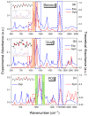
Figure 1
Figure 1
The comparison of the experimental spectra with the theoretical one (B3-LYP/6-311G (d,p)) for the synthetic absorbable threads in the region of 500–3000 cm−1. (a) Poly(glycolide/ε-caprolactone) copolymer (Monocryl); (b) Poly(glycolide/L-lactide) copolymer (Vicryl); (c) Poly-p-dioxanone. The blue dashed line shows the ester group, and red represents the ether group. Orange and green highlights show the characteristic bands for glycolide and dioxanone building blocks, respectively.
Figure 2
Figure 2
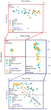
Principal component analysis (PCA) of the vector-normalized second-derivative FTIR spectra from the 950–1800 cm−1 region. The analysis encompasses data for various polymers across different environments and time points. Initially, PCA was conducted on the entire dataset, which was subsequently subdivided into two distinct panels to more clearly illustrate the differences between samples. The red-dashed rectangle highlights data specific to PDS and PDS Plus, while the blue-dashed rectangle encompasses data for Monocryl, Monocryl Plus, Vicryl, and Vicryl Plus.
Figure 3
Figure 3
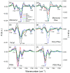
FTIR spectra of the fingerprint range of the hydrolytic bonds (ester and ether groups) for absorbable threads immersed in the sterile body fluids for 21 days. (a) Monocryl; (b) Vicryl; (c) PDS; (d) Monocryl Plus; (e) Vicryl Plus; (f) PDS plus. Black solid line—raw threads (before degradation); red line—saline solution; blue line—pancreatic juice; green line—bile.
Figure 4
Figure 4
FTIR spectra of characteristic ester bands sensitive to degradation under bacterial contamination after 21 days. (a) Monocryl; (b) Vicryl; (c) PDS; (d) Monocryl Plus; (e) Vicryl Plus; (f) PDS plus. Black solid line—raw thread (before degradation); blue line—pancreatic juice; green line—bile.
Figure 5
Figure 5
Position of bands on the spectra of raw polymers and after 21 days of incubation in body fluids within the range of 1700–1800 cm−1 (stretching vibration of the C=O group), determined on the basis of second derivatives. Smoothing was performed using a Savitsky–Golay filter with a score of 11. (ac) Uncoated surgical threads; (df) Triclosan-coated threads. Results for both sterile and contaminated environments.
Figure 6
Figure 6
Average percentage change in tensile strength and ester band absorbance for Monocryl/Monocryl plus threads relative to the reference (raw suture before degradation). (a,c) Sterile environment; (b,d) Contaminated environment. Top figures—strength results; bottom figures—FTIR results (ν C=O vibration). Light orange—7 days; light green—14 days; light cyan—21 days. The absorbance error is ±2 cm−1. Patterned columns represent coated sutures.
Figure 7
Figure 7
Average percentage change in tensile strength and ester band absorbance for Vicryl/Vicryl plus threads relative to the reference (raw suture before degradation). (a,c) Sterile environment; (b,d) Contaminated environment. Top figures—strength results; bottom figures—FTIR results (ν C=O vibration). Light orange—7 days; light green—14 days; light cyan—21 days; light gray—28 days. The absorbance error is ±2 cm−1. Patterned columns represent coated sutures.
Figure 8
Figure 8
Average percentage change in tensile strength and ester band absorbance for PDS/PDS plus threads relative to the reference (raw suture before degradation). (a,c) Sterile environment; (b,d) Contaminated environment. Top figures—strength results; bottom figures—FTIR results (ν C=O vibration). Light orange—7 days; light green—14 days; light cyan—21 days; light gray—28 days. The absorbance error is ±2 cm−1. Patterned columns represent coated sutures.
Figure 9
Figure 9
Scanning electron micrographs of uncoated threads immersed for 21 days in sterile saline, pancreatic, and bile juices. (a1a3) Saline; (b1b3) pancreatic juice; (c1c3) bile; Subscripts: 1- Monocryl, 2 -Vicryl, 3 -PDS threads.
Figure 10
Figure 10
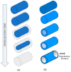
Mechanism of degradation of absorbable polymers: Surface erosion—observed for the Monocryl and the PDS threads (a). Bulk degradation—observed for the Vicryl threads (b).
Figure 11
Figure 11
DSC thermograms of fresh surgical threads made from PDS and stored in bile and pancreatic juice for 21 days in a contaminated environment.
Figure 12
Figure 12
Chemical structure of the polymers from which surgical threads are made. (a) Poly(glycolide/ε-caprolactone) Copolymer or Poliglecaprone 25. Suture trade name is Monocryl. (b) Poly(glycolide/L-lactide) Copolymer or Polyglactin 910 (m = 90, n = 10). Suture trade name is Vicryl. (c) Poly-p-dioxanone. Suture trade name is PDS II. (d) Antibacterial coating made of triclosan.

References

    1. Schneider L.A., Korber A., Grabbe S., Dissemond J. Influence of pH on wound-healing: A new perspective for wound-therapy? Arch. Dermatol. Res. 2007;298:413–420. doi: 10.1007/s00403-006-0713-x. - DOI - PubMed
    1. Adanur S. Wellington Sears Handbook of Industrial Textiles. 1st ed. CRC Press; Boca Raton, FL, USA: 1995. 832p
    1. Kim J.C., Lee Y.K., Lim B.S., Rhee S.H., Yang H.C. Comparison of tensile and knot security properties of surgical threads. J. Mater. Sci.-Mater. Med. 2007;18:2363–2369. doi: 10.1007/s10856-007-3114-6. - DOI - PubMed
    1. Nobile L., Checchi L., Monaco G. Experimental analysis of tensile properties of some suturing materials. J. Mater. Sci.-Mater. Med. 1997;8:53–56. doi: 10.1023/A:1018594330075. - DOI - PubMed
    1. Dennis C., Sethu S., Nayak S., Mohan L., Morsi Y., Manivasagam G. Suture materials—Current and emerging trends. J. Biomed. Mater. Res. A. 2016;104:1544–1559. doi: 10.1002/jbm.a.35683. - DOI - PubMed

LinkOut - more resources