. 2024 Oct 2;12(10):2243.
doi: 10.3390/biomedicines12102243.

Preconditioning of Mesenchymal Stem Cells Enhances the Neuroprotective Effects of Their Conditioned Medium in an Alzheimer's Disease In Vitro Model

Affiliations

Preconditioning of Mesenchymal Stem Cells Enhances the Neuroprotective Effects of Their Conditioned Medium in an Alzheimer's Disease In Vitro Model

Tatiana Tolstova et al. Biomedicines. .

Abstract

Background: Alzheimer's disease (AD) develops as a result of oxidative damage to neurons and chronic inflammation of microglia. These processes can be influenced by the use of a conditioned medium (CM) derived from mesenchymal stem cells (MSCs). The CM contains a wide range of factors that have neurotrophic, antioxidant, and anti-inflammatory effects. In addition, the therapeutic potential of the CM can be further enhanced by pretreating the MSCs to increase their paracrine activity. The current study aimed to investigate the neuroprotective effects of CM derived from MSCs, which were either activated by a TLR3 ligand or exposed to CoCl2, a hypoxia mimetic (pCM or hCM, respectively), in an in vitro model of AD.

Methods: We have developed a novel in vitro model of AD that allows us to investigate the neuroprotective and anti-inflammatory effects of MSCs on induced neurodegeneration in the PC12 cell line and the activation of microglia using THP-1 cells.

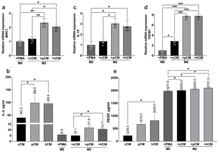
Results: This study demonstrates for the first time that pCM and hCM exhibit more pronounced immunosuppressive effects on proinflammatory M1 macrophages compared to CM derived from untreated MSCs (cCM). This may help prevent the development of neuroinflammation by balancing the M1 and M2 microglial phenotypes via the decreased secretion of proinflammatory cytokines (IL-1β, IL-6, and TNF-α) and increased secretion of IL-4, as well as the expression of IL-10 and TGF-β by macrophages. Moreover, a previously unknown increase in the neurotrophic properties of hCM was discovered, which led to an increase in the viability of neuron-like PC12 cells under H2O2-induced oxidative-stress conditions. These results are likely associated with an increase in the production of growth factors, including vascular endothelial growth factor (VEGF). In addition, the neuroprotective effects of CM from preconditioned MSCs are also mediated by the activation of the Nrf2/ARE pathway in PC12 cells.

Conclusions: TLR3 activation in MSCs leads to more potent immunosuppressive effects of the CM against pro-inflammatory M1 macrophages, while the use of hCM led to increased neurotrophic effects after H2O2-induced damage to neuronal cells. These results are of interest for the potential treatment of AD with CM from preactivated MSCs.

Keywords: Alzheimer’s disease; MSCs; PC12; immunosuppression; microglia; neuroprotection; secretome.


Conflict of interest statement

The authors declare no conflicts of interest. The funders had no role in the design of the study; in the collection, analyses, or interpretation of data; in the writing of the manuscript; or in the decision to publish the results.

Figures

Figure 1
Figure 1
NGF-induced neuronal differentiation of PC12 cells and the PMA-induced differentiation of THP-1 cells into macrophages. Investigation of morphology (a), neurite length (b,d), and relative number of differentiated PC12 cells (c). The neurites are highlighted in blue (b). Micrographs of THP-1 cells without PMA treatment (e), after PMA treatment (f) (10 ng/mL, 24 h), and after LPS-induced differentiation into the M1 phenotype (g) (10 ng/mL PMA, 24 h + 1 μg/mL LPS, 72 h). Scale bars, 50 μm (a,b) or 100 μm (eg). The error is indicated as SEM. * p < 0.05, ** p < 0.01, and *** p < 0.001.
Figure 2
Figure 2
Phenotypic characterization and expression of immunosuppressive molecules in human adipose-derived MSCs. (a) The fibroblast-like morphology of the adhesive cells was examined via phase-contrast microscopy. (b) The multipotency of the cells was confirmed through the results of cytochemical staining for calcium deposition (Alizarin Red) and lipid accumulation (Oil Red O) after the induction of osteogenesis and adipogenesis, respectively. Scale bar, 100 μm. The immunophenotypic characteristics of the MSCs were investigated via flow cytometry. Negative expression of the CD34, CD45, and HLA-DR markers was observed, whereas positive expression of the CD73, CD90, and CD105 markers was detected (c). The relative mRNA expression of the IDO1, TNFAIP6, and PTGES2 genes was also analyzed in hypoxia mimetic preconditioned MSCs (100 μM CoCl2 for 3 h) (d). Error bars indicate SEM. ** p < 0.01 and *** p < 0.001.
Figure 3
Figure 3
Investigation of the neurotrophic potential of CMs derived from MSCs. Morphology (a), neurite length (b), and number of differentiated PC12 cells (c) without treatment and after treatment with cCM, pCM, or hCM for 7 days. Scale bar, 50 μm. Neurites are highlighted in blue. Error bars indicate SEM. * p < 0.05.
Figure 4
Figure 4
Study of the neuroprotective properties of CMs derived from MSCs. Evaluation of cell viability (a), the expression of the BCL2 gene mRNA (b), the ROS level (c,d), and the mRNA expression of the NQO1 and HMOX1 genes (e) in differentiated PC12 cells without pretreatment (C, control group) and with pretreatment with different types of CM (cCM, pCM, and hCM) following H2O2-induced oxidative stress. Positive staining for DCF is shown in green, while cell nuclei are stained with Hoechst-33342 in blue. Scale bar, 50 µm (c). ROS production is expressed as a percentage (%) of DCF-stained cells relative to all cells (d). Error bars indicate SEM. * p < 0.05, ** p < 0.01, and *** p < 0.001.
Figure 5
Figure 5
Effect of CM derived from MSCs on the LPS-induced differentiation of macrophages from the M0 phenotype to the M1 phenotype. The levels of the proinflammatory cytokines TNF-α, IL-1β, and IL-6 secreted by MSCs and macrophages in the M0 and M1 states. Error bars indicate SEM. n.d.—not detected. * p < 0.05, ** p < 0.01, and *** p < 0.001.
Figure 6
Figure 6
The effect of MSC-derived CM on the polarization of M0 macrophages toward the M2 phenotype. (a,c,d) Relative expression levels of MRC1, IL-10, and TGFB1, respectively. (b,e) Secretion levels of IL-4 and VEGF, respectively, following the induction of differentiation with CMs. Error bars indicate SEM. * p < 0.05, ** p < 0.01, and *** p < 0.001.

References

    1. Alidoust L., Akhoondian M., Atefi A.H., Keivanlou M.-H., Hedayati Ch M., Jafari A. Stem cell-conditioned medium is a promising treatment for Alzheimer’s disease. Behav. Brain Res. 2023;452:114543. doi: 10.1016/j.bbr.2023.114543. - DOI - PubMed
    1. Rosini M., Simoni E., Milelli A., Minarini A., Melchiorre C. Oxidative Stress in Alzheimer’s Disease: Are We Connecting the Dots?: Miniperspective. J. Med. Chem. 2014;57:2821–2831. doi: 10.1021/jm400970m. - DOI - PubMed
    1. Kim J., Lee Y., Lee S., Kim K., Song M., Lee J. Mesenchymal Stem Cell Therapy and Alzheimer’s Disease: Current Status and Future Perspectives. J. Alzheimer’s Dis. 2020;77:1–14. doi: 10.3233/JAD-200219. - DOI - PubMed
    1. Yokokawa K., Iwahara N., Hisahara S., Emoto M.C., Saito T., Suzuki H., Manabe T., Matsumura A., Matsushita T., Suzuki S., et al. Transplantation of Mesenchymal Stem Cells Improves Amyloid-β Pathology by Modifying Microglial Function and Suppressing Oxidative Stress. J. Alzheimer’s Dis. 2019;72:867–884. doi: 10.3233/JAD-190817. - DOI - PMC - PubMed
    1. Shin J.Y., Park H.J., Kim H.N., Oh S.H., Bae J.-S., Ha H.-J., Lee P.H. Mesenchymal stem cells enhance autophagy and increase β-amyloid clearance in Alzheimer disease models. Autophagy. 2014;10:32–44. doi: 10.4161/auto.26508. - DOI - PMC - PubMed

LinkOut - more resources