. 2024 Oct 10;12(10):2299.
doi: 10.3390/biomedicines12102299.

Protective Effects of Trimetazidine and Dexmedetomidine on Liver Injury in a Mesenteric Artery Ischemia-Reperfusion Rat Model via Endoplasmic Reticulum Stress

Affiliations

Protective Effects of Trimetazidine and Dexmedetomidine on Liver Injury in a Mesenteric Artery Ischemia-Reperfusion Rat Model via Endoplasmic Reticulum Stress

Sedat Ciftel et al. Biomedicines. .

Abstract

Background/objectives: Acute mesenteric ischemia can lead to severe liver damage due to ischemia-reperfusion (I/R) injury. This study investigated the protective effects of trimetazidine (TMZ) and dexmedetomidine (DEX) against liver damage induced by mesenteric artery I/R via endoplasmic reticulum stress (ERS) mechanisms.

Methods: Twenty-four rats were divided into four groups: control, I/R, I/R+TMZ, and I/R+DEX. TMZ (20 mg/kg) was administered orally for seven days, and DEX (100 µg/kg) was given intraper-itoneally 30 min before I/R induction. Liver tissues were analyzed for creatinine, alanine ami-notransferase (ALT), aspartate aminotransferase (AST), thiobarbituric acid reactive substances (TBARS), and total thiol (TT) levels.

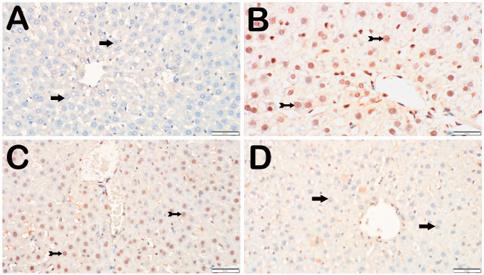
Results: Compared with the control group, the I/R group presented significantly increased AST, ALT, TBARS, and TT levels. TMZ notably reduced creatinine levels. I/R caused significant liver necrosis, inflammation, and congestion. TMZ and DEX treatments reduced this histopathological damage, with DEX resulting in a more significant reduction in infiltrative areas and vascular congestion. The increase in the expression of caspase-3, Bax, 8-OHdG, C/EBP homologous protein (CHOP), and glucose-regulated protein 78 (GRP78) decreased with the TMZ and DEX treatments. In addition, Bcl-2 positivity decreased both in the TMZ and DEX treatments.

Conclusions: Both TMZ and DEX have protective effects against liver damage. These effects are likely mediated through the reduction in ERS and apoptosis, with DEX showing slightly superior protective effects compared with TMZ.

Keywords: dexmedetomidine; endoplasmic reticulum stress; ischemia–reperfusion injury; liver; mesenteric artery ischemia; trimetazidine.


Conflict of interest statement

The authors declare that they have no known competing financial interests or personal relationships that could have appeared to influence the work reported in this paper.

Figures

Figure 1
Figure 1
Representative light microscopic screen image of H&E-stained liver tissue sections. Central vein (CV). (A) (×20) and (B) (×40). Control group: In the liver tissue sections belonging to the control group, Remark cords containing hepatocytes with a normal structure (arrow) were observed (LHDS: 0(0-1)). (C) (×20) and (D) (×40). I/R group: In the liver tissue, widespread necrotic hepatocytes (arrowhead) are observed, primarily in Zone 1 (Z1) of the intralobular areas. Additionally, necrotic hepatocytes (arrowhead), vascular congestion (asteriks), and infiltrative areas (tailed arrow) are observed in the perilobular regions. (LHDS: 9(8-10)). (E) (×20) and (F) (×40). I/R+TMZ group: In the liver tissue, a reduction in intralobular necrosis, particularly perilobular necrosis, was observed. Additionally, a decrease in periportal infiltrative areas and vascular congestion was noted (LHDS: 4(3-5)). (G) (×20) and (H) (×40). I/R+DEX group: In the liver tissue, a decrease in necrotic hepatocytes was observed in both the intralobular and perilobular areas. Additionally, a reduction in periportal infiltrative areas and vascular congestion was noted (LHDS: 2(1-3)).
Figure 2
Figure 2
Representative light microscopic screen images of liver tissue sections incubated with a Caspase-3 primary antibody. (A) (×20). Control group: In the liver tissue sections from the control group, hepatocytes with a normal structure (arrow) were immunonegative (caspase-3 positivity score: 0(0-0)). (B) (×20). I/R group: In the liver tissue, hepatocytes showing intense Caspase-3 immunopositivity (spiral arrow) are observed, primarily in Zone 1 of the intralobular areas (Caspase-3 positivity score: 1(1-2)). (C) (×20). I/R+TMZ group: In the liver tissue, a decrease in the number of hepatocytes positive for Caspase-3 was observed, particularly in the intralobular and perilobular areas (Caspase-3 positivity score: 0.5(0-1)). (D) (×20). I/R+DEX group: A decrease in the number of apoptotic hepatocytes positive for Caspase-3 in the intralobular and perilobular areas was observed, with a widespread presence of immunonegative hepatocytes (caspase-3 positivity score: 0(0-1)).
Figure 3
Figure 3
Representative light microscopic screen images of liver tissue sections incubated with 8-OHdG primary antibody. (A) (×20). Control group: In the liver tissue sections from the control group, hepatocytes with a normal structure (arrow) were 8-OHdG-negative (8-OHdG positivity score: 0(0-0)). (B) (×20). I/R group: In the liver tissue, hepatocytes showing intense immunopositivity with 8-OHdG primary antibody (spiral arrow) are observed, primarily in Zone 1 of the intralobular areas (8-OHdG positivity score: 2(2-3)). (C) (×20). I/R+TMZ group: A decrease in hepatocytes showing 8-OHdG immunopositivity was observed in the intralobular and perilobular areas (8-OHdG positivity score: 1(1-2)). (D) (×20). I/R+DEX group: A decrease in hepatocytes showing intense 8-OHdG immunopositivity in the intralobular and perilobular areas was observed, with a widespread presence of immunonegative hepatocytes (arrow, 8-OHdG positivity score: 1(0-1)).
Figure 4
Figure 4
Representative light microscopic screen images of liver tissue sections incubated with CHOP primary antibody. (A) (×20). Control group: In the liver tissue sections from the control group, hepatocytes with a normal structure (arrow) were observed to be CHOP-negative (CHOP positivity score: 0(0-0)). (B) (×20). I/R group: In the intralobular areas, primarily in Zone 1, hepatocytes showing intense CHOP positivity (spiral arrow) are observed (CHOP positivity score: 2(1-2)). (C) (×20). I/R+TMZ group: A decrease in hepatocytes showing CHOP positivity was observed in the perinobular areas, particularly in the intralobular regions (CHOP positivity score: 1(0-1)). (D) (×20). I/R+DEX group: A decrease in hepatocytes showing intense immunopositivity in the intralobular and perilobular areas was observed, with a widespread presence of CHOP-negative hepatocytes (arrow, CHOP positivity score: 0.5 (0-1)).
Figure 5
Figure 5
Representative light microscopy images of liver tissue sections incubated with an anti-GRP78 primary antibody. (A) (×20). Control group: In the liver tissue sections from the control group, hepatocytes with a normal structure (arrow) were observed to be GRP78-negative (GRP78 positivity score: 0(0-0)). (B) (×20). I/R group: In the intralobular areas, primarily in Zone 1, hepatocytes showing intense GRP78 positivity (spiral arrow) are observed (GRP78 positivity score: 2(2-2)). (C) (×20). I/R+TMZ group: A decrease in hepatocytes showing GRP78 positivity was observed in the perilobular areas, particularly in the intralobular regions (GRP78 positivity score: 1.5(1-2)). (D) (×20). I/R+DEX group: A decrease in hepatocytes showing intense immunopositivity in the intralobular and perilobular areas was observed, with a widespread presence of GRP78-negative hepatocytes (arrow, GRP78 positivity score: 1(0-1)).
Figure 6
Figure 6
Representative light microscopy images of liver tissue sections incubated with a Bax primary antibody. (A) (×20). Control group: In the liver tissue sections from the control group, hepatocytes with a normal structure (arrow) were observed to be Bax-negative (Bax positivity score: 0(0-0)). (B) (×20). I/R group: In the intralobular areas, which are primarily in Zone 1, hepatocytes showing intense Bax positivity (spiral arrow) are observed (Bax positivity score: 2(2-3)). (C) (×20). I/R+TMZ group: A decrease in hepatocytes showing Bax positivity was observed in the perilobular areas, particularly in the intralobular regions (Bax positivity score: 1(1-1.5)). (D) (×20). I/R+DEX group: A decrease in hepatocytes showing intense immunopositivity in the intralobular and perilobular areas was observed, with a widespread presence of Bax-negative hepatocytes (arrow, Bax positivity score: 0(0-1)).
Figure 7
Figure 7
Representative light microscopy images of liver tissue sections incubated with a Bcl-2 primary antibody. (A) (×20). Control group: In the liver tissue sections from the control group, hepatocytes with a normal structure (arrow) were observed to be Bcl-2-negative (Bcl-2 positivity score: 0(0-0)). (B) (×20). I/R group: In the intralobular areas, primarily in Zone 1, hepatocytes showing Bcl-2 negativity (spiral arrow) are observed (Bcl-2 positivity score: 0(0-1)). (C) (×20). I/R+TMZ group: An increase in hepatocytes showing Bcl-2 positivity was observed in the perilobular areas, particularly in the intralobular regions (Bcl-2 positivity score: 1(1-2)). (D) (×20). I/R+DEX group: An increase in hepatocytes showing intense immunopositivity in the intralobular and perilobular areas was observed, with a widespread presence of Bcl-2-positive hepatocytes (arrow, Bcl-2 positivity score: 2(2-3)).

Similar articles

References

    1. Liu J., Pan G., Liang T., Huang P. HGF/c-Met Signaling Mediated Mesenchymal Stem Cell-Induced Liver Recovery in Intestinal Ischemia Reperfusion Model. Int. J. Med. Sci. 2014;11:626–633. doi: 10.7150/ijms.8228. - DOI - PMC - PubMed
    1. Zhao J., Chen X.D., Yan Z.Z., Huang W.F., Liu K.X., Li C. Gut-Derived Exosomes Induce Liver Injury after Intestinal Ischemia/Reperfusion by Promoting Hepatic Macrophage Polarization. Inflammation. 2022;45:2325–2338. doi: 10.1007/s10753-022-01695-0. - DOI - PubMed
    1. Konishi T., Lentsch A.B. Hepatic Ischemia/Reperfusion: Mechanisms of Tissue Injury, Repair, and Regeneration. Gene Expr. 2017;17:277–287. doi: 10.3727/105221617X15042750874156. - DOI - PMC - PubMed
    1. Sharp A.J., Patel N., Reeves B.C., Angelini G.D., Fiorentino F. Pharmacological Interventions for the Prevention of Contrast-Induced Acute Kidney Injury in High-Risk Adult Patients Undergoing Coronary Angiography: A Systematic Review and Meta-Analysis of Randomized Controlled Trials. Open Heart. 2019;6:e000864. doi: 10.1136/openhrt-2018-000864. - DOI - PMC - PubMed
    1. Mahfoudh-Boussaid A., Tka K.H.A., Zaouali M.A., Roselló-Catafau J., Ben Abdennebi H. Effects of Trimetazidine on the Akt/ENOS Signaling Pathway and Oxidative Stress in an in Vivo Rat Model of Renal Ischemia–Reperfusion. Ren. Fail. 2014;36:1436–1442. doi: 10.3109/0886022X.2014.949765. - DOI - PubMed

LinkOut - more resources