

A Transfer Learning-Based Framework for Classifying Lymph Node Metastasis in Prostate Cancer Patients
- PMID: 39457657
- PMCID: PMC11504638
- DOI: 10.3390/biomedicines12102345
A Transfer Learning-Based Framework for Classifying Lymph Node Metastasis in Prostate Cancer Patients
Abstract
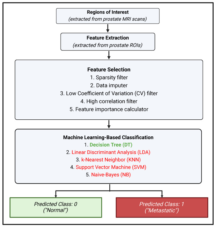
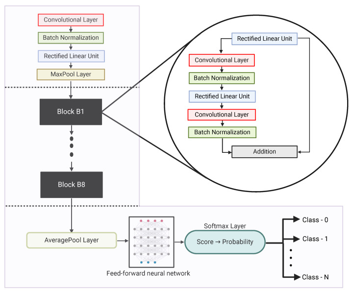
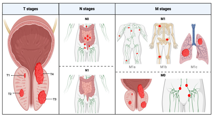
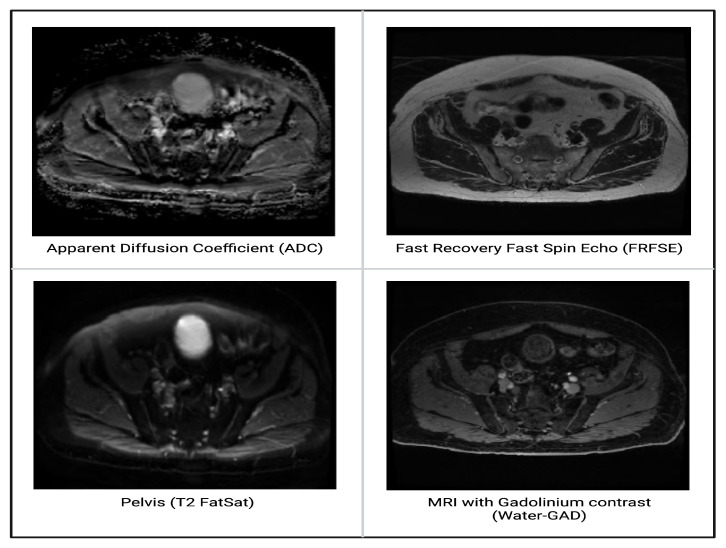
Background: Prostate cancer is the second most common new cancer diagnosis in the United States. It is usually slow-growing, and when it is low-grade and confined to the prostate gland, it can be treated either conservatively (through active surveillance) or with surgery. However, if the cancer has spread beyond the prostate, such as to the lymph nodes, then that indicates a more aggressive cancer, and surgery may not be adequate. Methods: The challenge is that it is often difficult for radiologists reading prostate-specific imaging such as magnetic resonance images (MRIs) to differentiate malignant lymph nodes from non-malignant ones. An emerging field is the development of artificial intelligence (AI) models, including machine learning and deep learning, for medical imaging to assist in diagnostic tasks. Earlier research focused on implementing texture algorithms to extract imaging features used in classification models. More recently, researchers began studying the use of deep learning for both stand-alone feature extraction and end-to-end classification tasks. In order to tackle the challenges inherent in small datasets, this study was designed as a scalable hybrid framework utilizing pre-trained ResNet-18, a deep learning model, to extract features that were subsequently fed into a machine learning classifier to automatically identify malignant lymph nodes in patients with prostate cancer. For comparison, two texture algorithms were implemented, namely the gray-level co-occurrence matrix (GLCM) and Gabor. Results: Using an institutional prostate lymph node dataset (42 positives, 84 negatives), the proposed framework achieved an accuracy of 76.19%, a sensitivity of 79.76%, and a specificity of 69.05%. Using GLCM features, the classification achieved an accuracy of 61.90%, a sensitivity of 74.07%, and a specificity of 42.86%. Using Gabor features, the classification achieved an accuracy of 65.08%, a sensitivity of 73.47%, and a specificity of 52.50%. Conclusions: Our results demonstrate that a hybrid approach, i.e., using a pre-trainined deep learning model for feature extraction, followed by a machine learning classifier, is a viable solution. This hybrid approach is especially useful in medical-imaging-based applications with small datasets.
Keywords: deep learning; lymph node metastasis; machine learning; magnetic resonance imaging; prostate cancer.
Conflict of interest statement
The authors declare no conflicts of interest.
Figures
References
-
- Key Statistics for Prostate Cancer—American Cancer Society. [(accessed on 28 September 2023)]. Available online: https://www.cancer.org/cancer/prostate-cancer/about/key-statistics.html.
-
- Prostate Cancer—Symptoms and Causes—Mayo Clinic. [(accessed on 28 September 2023)]. Available online: https://www.mayoclinic.org/diseases-conditions/prostate-cancer/symptoms-....
-
- Ghaderzadeh M. Clinical Decision Support System for Early Detection of Prostate Cancer from Benign Hyperplasia of Prostate. MEDINFO, IOS Press; Amsterdam, The Netherlands: 2013. p. 928. - PubMed
-
- Sadoughi F., Ghaderzadeh M. A Hybrid Particle Swarm and Neural Network Approach for Detection of Prostate Cancer from Benign Hyperplasia of Prostate. IOS Press; Amsterdam, The Netherlands: 2014. pp. 481–485. InE-Health–For Continuity of Care. - PubMed
Grants and funding
LinkOut - more resources
Full Text Sources
