Review
. 2024 Oct 15;12(10):2346.
doi: 10.3390/biomedicines12102346.

The Role of Immune Cells and Signaling Pathways in Diabetic Eye Disease: A Comprehensive Review

Affiliations
Review

The Role of Immune Cells and Signaling Pathways in Diabetic Eye Disease: A Comprehensive Review

Vincenzo Barone et al. Biomedicines. .

Abstract

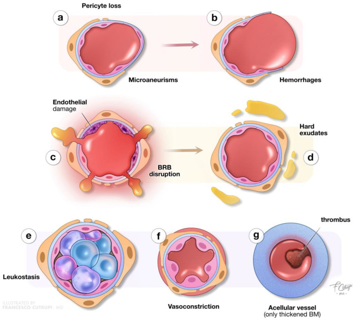
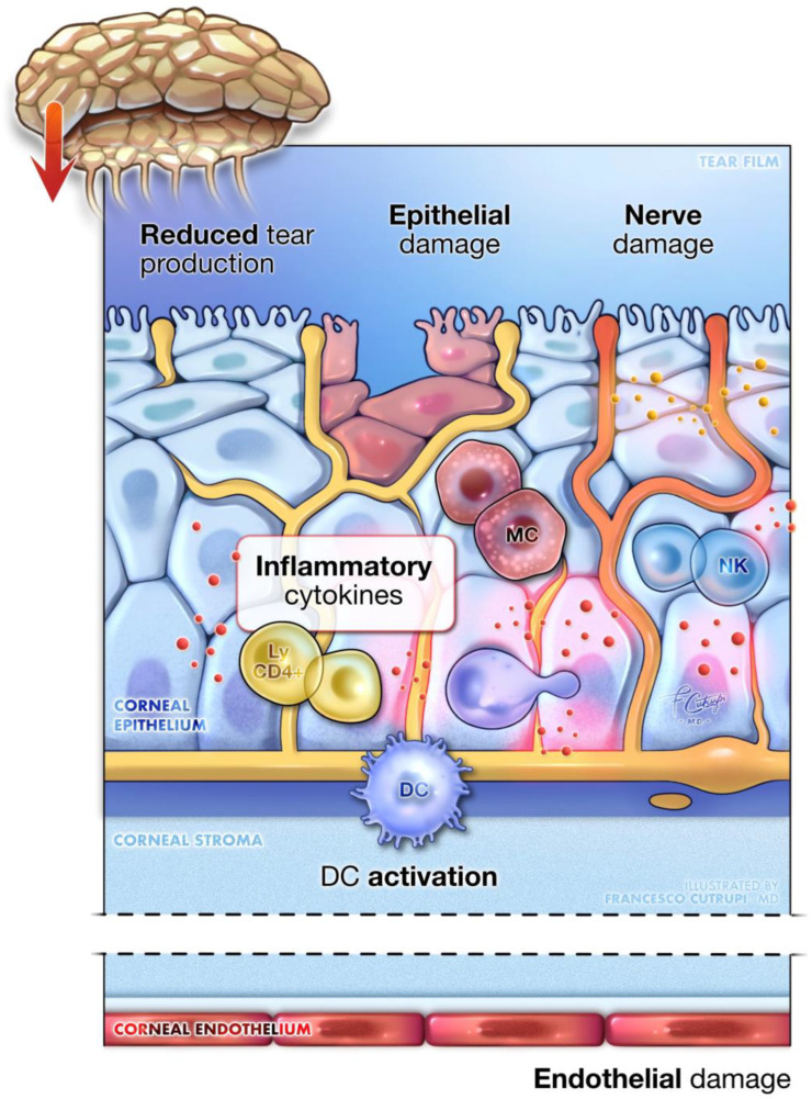
Diabetic eye disease (DED) encompasses a range of ocular complications arising from diabetes mellitus, including diabetic retinopathy, diabetic macular edema, diabetic keratopathy, diabetic cataract, and glaucoma. These conditions are leading causes of visual impairments and blindness, especially among working-age adults. Despite advancements in our understanding of DED, its underlying pathophysiological mechanisms remain incompletely understood. Chronic hyperglycemia, oxidative stress, inflammation, and neurodegeneration play central roles in the development and progression of DED, with immune-mediated processes increasingly recognized as key contributors. This review provides a comprehensive examination of the complex interactions between immune cells, inflammatory mediators, and signaling pathways implicated in the pathogenesis of DED. By delving in current research, this review aims to identify potential therapeutic targets, suggesting directions of research for future studies to address the immunopathological aspects of DED.

Keywords: diabetes; diabetic eye disease; diabetic keratopathy; diabetic retinopathy; immune cells; inflammation; pathophysiology; signaling pathways.


Conflict of interest statement

The authors declare no conflicts of interest related to this study.

Figures

Figure 1
Figure 1
Clinical manifestations of diabetic eye disease. This illustration provides a comprehensive overview of the ocular signs associated with diabetes mellitus. It highlights key manifestations including corneal damage, cataract formation, retinal degeneration, and the development of glaucoma (IOP: intraocular pressure; IRMAs: intraretinal microvascular abnormalities).
Figure 2
Figure 2
Microvascular alterations in non-proliferative diabetic retinopathy. This illustration details the microvascular changes occurring in non-proliferative diabetic retinopathy. Chronic hyperglycemia results in the loss of pericytes in retinal capillaries, undermining structural support and leading to the formation of microaneurysms (a) and vessel ruptures accompanied by hemorrhages (b). Strong inflammatory processes, along with endothelial and basement membrane (BM) alterations, compromise the blood–retinal barrier (BRB), increasing vascular permeability (c) and resulting in edema and hard exudates (d). Furthermore, the retinal blood flow is strongly compromised due to leukostasis (with vascular occlusion), vasoconstriction, and the presence of highly thrombogenic acellular capillaries. Furthermore, the retinal-blood-flow is-strongly-compromised due to leukostasis (with-vascular occlusion) (e), vasoconstriction (f), and the presence of-highly-thrombo-genic acellular capillaries (g).
Figure 3
Figure 3
Diabetic keratopathy. A detailed close-up of corneal tissue showcases the main pathogenic mechanisms underlying diabetic keratopathy. The image illustrates the epithelial dysfunction seen, alongside pronounced inflammation, mediated by immune cells within the cornea. Additionally, it depicts damage to the corneal nerves and lacrimal gland innervation, leading to decreased sensitivity, neurotrophic alterations, and reduced tear production. Endothelial cell damage is also present, leading to corneal edema and thickening. Key inflammatory cells include the activation of dendritic cells (DC), mast cells (MC), natural killer lymphocytes (NK), and resident memory CD4⁺ lymphocytes (ly CD4⁺).

Similar articles

Cited by

References

    1. American Diabetes Association Diagnosis and Classification of Diabetes Mellitus. Diabetes Care. 2014;37((Suppl. S1)):S81–S90. doi: 10.2337/dc14-S081. - DOI - PubMed
    1. Sun H., Saeedi P., Karuranga S., Pinkepank M., Ogurtsova K., Duncan B.B., Stein C., Basit A., Chan J.C.N., Mbanya J.C., et al. IDF Diabetes Atlas: Global, Regional and Country-Level Diabetes Prevalence Estimates for 2021 and Projections for 2045. Diabetes Res. Clin. Pract. 2022;183:109119. doi: 10.1016/j.diabres.2021.109119. - DOI - PMC - PubMed
    1. Mesquida M., Drawnel F., Fauser S. The Role of Inflammation in Diabetic Eye Disease. Semin. Immunopathol. 2019;41:427–445. doi: 10.1007/s00281-019-00750-7. - DOI - PubMed
    1. Bourne R.R.A., Jonas J.B., Bron A.M., Cicinelli M.V., Das A., Flaxman S.R., Friedman D.S., Keeffe J.E., Kempen J.H., Leasher J., et al. Prevalence and Causes of Vision Loss in High-Income Countries and in Eastern and Central Europe in 2015: Magnitude, Temporal Trends and Projections. Br. J. Ophthalmol. 2018;102:575–585. doi: 10.1136/bjophthalmol-2017-311258. - DOI - PMC - PubMed
    1. Li J.Q., Welchowski T., Schmid M., Letow J., Wolpers C., Pascual-Camps I., Holz F.G., Finger R.P. Prevalence, Incidence and Future Projection of Diabetic Eye Disease in Europe: A Systematic Review and Meta-Analysis. Eur. J. Epidemiol. 2020;35:11–23. doi: 10.1007/s10654-019-00560-z. - DOI - PubMed

LinkOut - more resources