. 2024 Oct 15;12(10):2347.
doi: 10.3390/biomedicines12102347.

Intestinal Region-Dependent Impact of NFκB-Nrf Crosstalk in Myenteric Neurons and Adjacent Muscle Cells in Type 1 Diabetic Rats

Affiliations

Intestinal Region-Dependent Impact of NFκB-Nrf Crosstalk in Myenteric Neurons and Adjacent Muscle Cells in Type 1 Diabetic Rats

Bence Pál Barta et al. Biomedicines. .

Abstract

Background/objectives: Type 1 diabetes affects cytokines as potential inducers of NFκB signalling involved in inflammation and neuronal survival. Our goal was to assess the expression of NFκB p65 and its negative regulator, Nrf2, in myenteric neurons and adjacent smooth muscle of different gut segments after chronic hyperglycaemia and immediate insulin treatment.

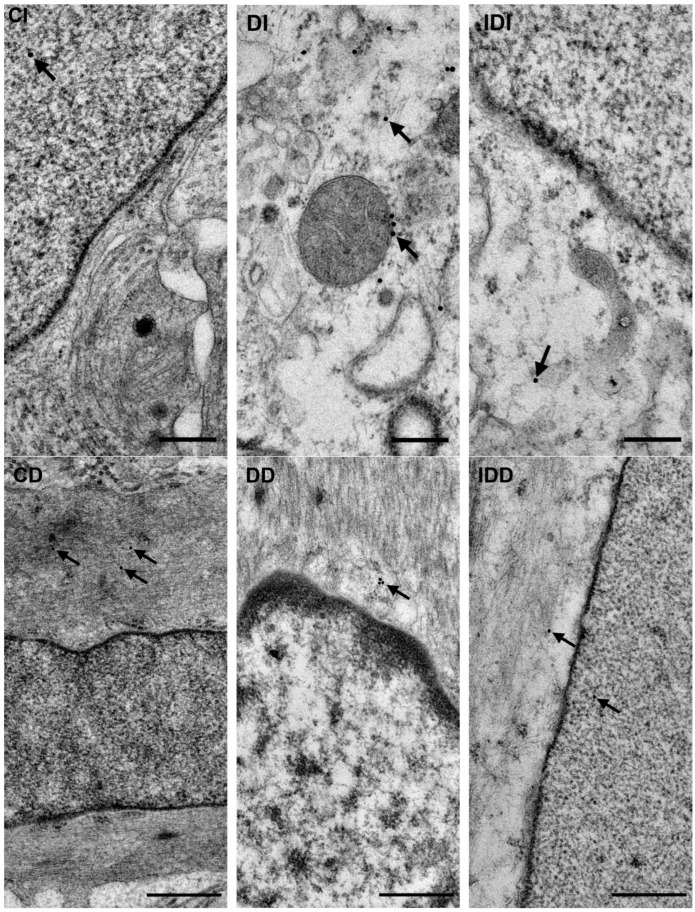
Methods: After ten weeks of hyperglycaemia, intestinal samples of control, streptozotocin-induced diabetic and insulin-treated diabetic rats were prepared for fluorescent immunohistochemistry, immunogold electron microscopy, ELISA and qPCR.

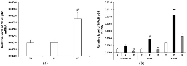
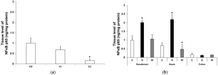
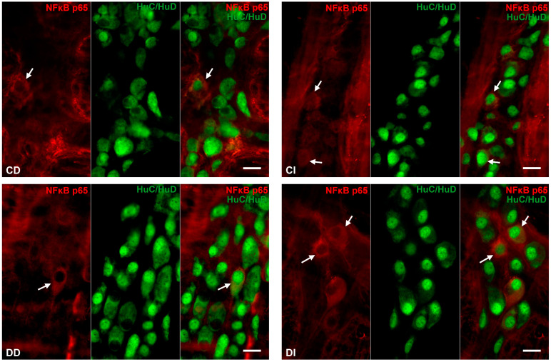
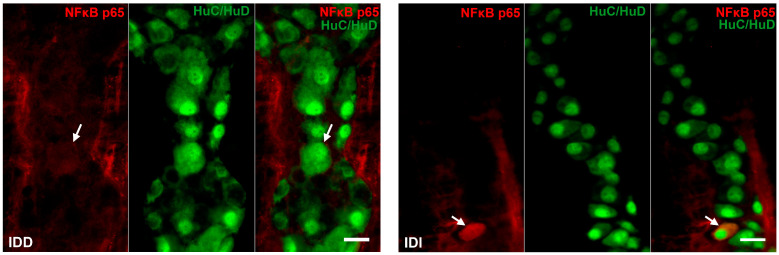
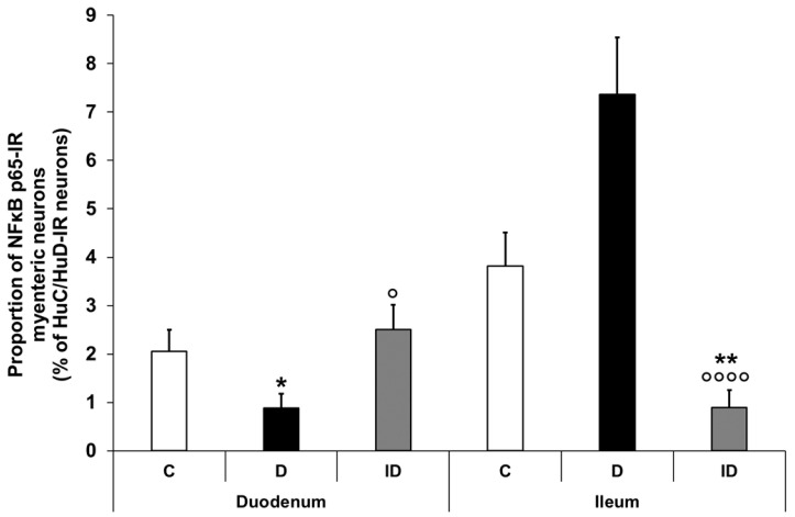
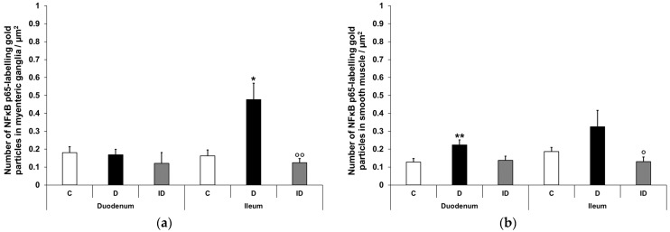
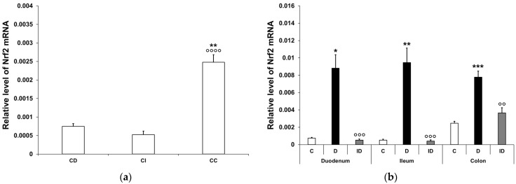
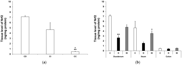
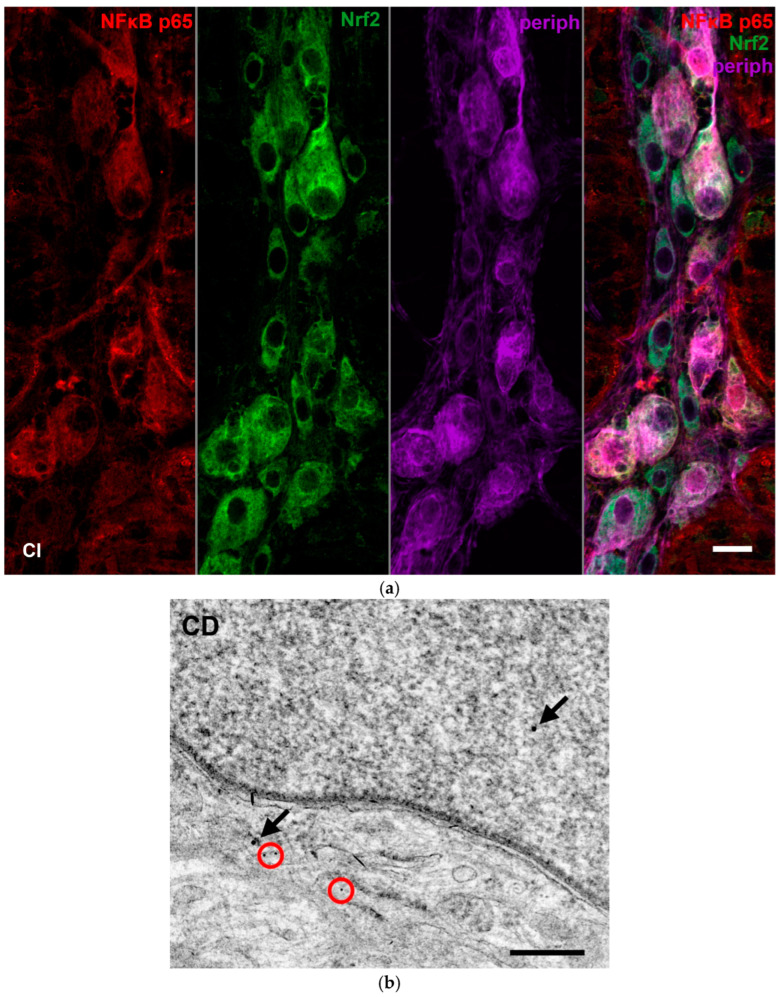
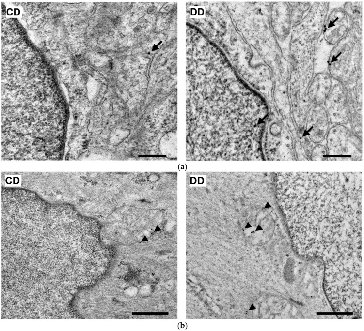
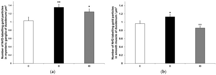
Results: In the diabetic rats, the proportion of NFκB p65-immunoreactive myenteric neurons decreased significantly in the duodenum and increased in the ileum. The density of NFκB p65-labelling gold particles increased in the ileal but remained unchanged in the duodenal ganglia. Meanwhile, both total and nuclear Nrf2 density increased in the myenteric neurons of the diabetic duodenum. In smooth muscle, NFκB p65 and Nrf2 density increased in the small intestine of diabetic rats. While on the mRNA level, NFκB p65 and Nrf2 were induced, on the protein level, NFκB p65 increased and Nrf2 decreased in muscle/myenteric plexus homogenates. Insulin treatment had protective effects.

Conclusions: Our findings reveal a segment-specific NFκB and Nrf expression in myenteric neurons and ganglionic muscular environments, which may contribute to regional neuronal survival and motility disturbances in diabetes.

Keywords: NFκB; Nrf2; animal model; enteric neurons; gut segments; hyperglycaemia; insulin; intestinal smooth muscle; myenteric plexus; type 1 diabetes.


Conflict of interest statement

The authors declare no conflicts of interest. The funders had no role in the design of the study; in the collection, analyses, or interpretation of data; in the writing of the manuscript; or in the decision to publish the results.

Figures

Figure 1
Figure 1
Relative levels of NFκB p65 mRNA. (a) Relative levels of NFκB p65 mRNA in tissue homogenates from different gut segments of control rats. The expression level of NFκB p65 mRNA was nearly three times higher in the colon than in the small intestinal segments of controls. Data were expressed as means ± SEM. ** p < 0.01 (compare with CD); oo p < 0.01 (between CI and CC). CD—control duodenum, CI—control ileum, CC—control colon. (b) Effects of long-lasting hyperglycaemia and insulin treatment on the relative level of NFκB p65 mRNA in tissue homogenates from different gut segments. In the diabetics, the relative level of NFκB p65 mRNA displayed a more than 3-fold increase in the tissue homogenates of the colon and ileum, which was prevented by insulin treatment. Data were expressed as means ± SEM. ** p < 0.01 (compare to C); oo p < 0.01 (between D and ID). C—controls, D—diabetics, ID—insulin-treated diabetics.
Figure 2
Figure 2
Tissue levels of NFκB p65. (a) Tissue levels of NFκB p65 in intestinal smooth muscle/myenteric plexus homogenates from different gut segments of control rats. The tissue level of NFκB p65 displayed a significant decrease from the duodenum to the colon of controls. Data were expressed as means ± SEM. * p < 0.05 (compare with CD). CD—control duodenum, CI—control ileum, CC—control colon. (b) Effects of long-lasting hyperglycaemia and insulin treatment on the tissue levels of NFκB p65 in smooth muscle/myenteric plexus homogenates from different gut segments. In the diabetic rats, the NFκB p65 level was doubled in the duodenum and tripled in the ileum, while it did not change in the colon. Immediate insulin treatment was completely protective against diabetes-related changes. Data were expressed as means ± SEM. * p < 0.05 (compare with C); o p < 0.05 (between D and ID). C—controls, D—diabetics, ID—insulin-treated diabetics.
Figure 3
Figure 3
Representative fluorescent micrographs of whole-mount preparations of myenteric ganglia from the duodenum and ileum of control, diabetic and insulin-treated diabetic rats after NFκB p65-HuC/HuD double-labelling immunohistochemistry. HuC/HuD as a pan-neuronal marker was applied to label myenteric neurons. CD—control duodenum, CI—control ileum, DD—diabetic duodenum, DI—diabetic ileum, IDD—insulin-treated diabetic duodenum, IDI—insulin-treated diabetic ileum, arrows—NFκB p65-immunoreactive myenteric neurons. Scale bars: 20 μm.
Figure 3
Figure 3
Representative fluorescent micrographs of whole-mount preparations of myenteric ganglia from the duodenum and ileum of control, diabetic and insulin-treated diabetic rats after NFκB p65-HuC/HuD double-labelling immunohistochemistry. HuC/HuD as a pan-neuronal marker was applied to label myenteric neurons. CD—control duodenum, CI—control ileum, DD—diabetic duodenum, DI—diabetic ileum, IDD—insulin-treated diabetic duodenum, IDI—insulin-treated diabetic ileum, arrows—NFκB p65-immunoreactive myenteric neurons. Scale bars: 20 μm.
Figure 4
Figure 4
Proportion of NFκB p65-immunoreactive myenteric neurons in the duodenum and ileum of control, diabetic and insulin-treated diabetic rats. In the diabetics, the proportion of NFκB p65-immunoreactive myenteric neurons was significantly decreased in the duodenum and increased in the ileum, which was prevented by immediate insulin treatment. Data were expressed as mean ± SEM. * p < 0.05, ** p < 0.01 (compare with C); o p < 0.05, oooo p < 0.0001 (between D and ID). C—controls, D—diabetics, ID—insulin-treated diabetics.
Figure 5
Figure 5
Representative electron micrographs of portions of the perikaryon and nuclei of myenteric neurons from ileum and intestinal smooth muscle cells from duodenum of control, diabetic and insulin-treated diabetic rats after NFκB p65 post-embedding immunohistochemistry. CD—control duodenum, CI—control ileum, DD—diabetic duodenum, DI—diabetic ileum, IDD—insulin-treated diabetic duodenum, IDI—insulin-treated diabetic ileum, arrows—18 nm gold particles’ labelling NFκB p65. Scale bars: 250 nm.
Figure 6
Figure 6
Quantification of gold particles’ labelling NFκB p65 in myenteric ganglia (a) and intestinal smooth muscle (b) from different gut segments of control, diabetic and insulin-treated diabetic rats. The number of NFκB p65-labelling gold particles increased in the ileal myenteric ganglia and intestinal smooth muscle of both the duodenum and the ileum of diabetic animals relative to the controls, which was prevented by insulin. Data were expressed as means ± SEM. * p < 0.05, ** p < 0.01 (compare with C); o p < 0.05, oo p < 0.01 (between D and ID). C—controls, D—diabetics, ID—insulin-treated diabetics.
Figure 7
Figure 7
Relative levels of Nrf2 mRNA. (a) Relative levels of Nrf2 mRNA in tissue homogenates from different gut segments of control rats. The expression level of Nrf2 mRNA was multiple times higher in the colon than in the small intestinal segments of the controls. Data were expressed as means ± SEM. ** p < 0.01 (compare with CD); oooo p < 0.0001 (between CI and CC). CD—control duodenum, CI—control ileum, CC—control colon. (b) Effects of long-lasting hyperglycaemia and insulin treatment on the relative level of Nrf2 mRNA in tissue homogenates from different gut segments. In the diabetics, the relative level of Nrf2 mRNA displayed a robust increase in all segments along the duodenum–ileum–colon axis, which was prevented by insulin treatment. Data were expressed as means ± SEM. * p < 0.05, ** p < 0.01, *** p < 0.001 (compare with C); oo p < 0.01, ooo p < 0.001 (between D and ID). C—controls, D—diabetics, ID—insulin-treated diabetics.
Figure 8
Figure 8
Tissue levels of Nrf2. (a) Tissue levels of Nrf2 in intestinal smooth muscle/myenteric plexus homogenates from different gut segments of control rats. The tissue levels of Nrf2 were significantly higher in the small intestine than the colon of the controls. Data were expressed as means ± SEM. ** p < 0.01 (compare with CD); o p < 0.05 (between CI and CC). CD—control duodenum, CI—control ileum, CC—control colon. (b) Effects of long-lasting hyperglycaemia and insulin treatment on the tissue levels of Nrf2 in smooth muscle/myenteric plexus homogenates from different gut segments. In the diabetic rats, the Nrf2 level was decreased in the duodenum and ileum, while it did not change in the colon. Immediate insulin treatment was protective against diabetes-related changes. Data were expressed as means ± SEM. ** p < 0.01 (compare with C); o p < 0.05 (between D and ID). C—controls, D—diabetics, ID—insulin-treated diabetics.
Figure 9
Figure 9
Representative fluorescent micrograph of a whole-mount preparation of myenteric ganglia from the ileum of a control rat after NFκB p65-Nrf2-Peripherin triple-labelling immunohistochemistry (a). Pan-neuronal Peripherin was used to label myenteric neurons. Representative electron micrograph of a portion of the perikaryon and nucleus of a myenteric neuron from duodenum of a control rat after NFκB p65-Nrf2 post-embedding immunohistochemistry (b). Red circles—10 nm gold particles’ labelling NFκB p65, arrows—18 nm gold particles’ labelling Nrf2. CI—control ileum, CD—control duodenum. Scale bars: 20 µm (a), 250 nm (b).
Figure 10
Figure 10
Representative electron micrographs of portions of the perikaryon and nuclei of myenteric neurons (a) and intestinal smooth muscle cells (b) from the duodenum of control and diabetic rats after Nrf2 post-embedding immunohistochemistry. CD—control duodenum, DD—diabetic duodenum. Arrows—18 nm gold particles’ labelling Nrf2 in myenteric neurons, arrowheads—18 nm gold particles’ labelling Nrf2 in muscle cells. Scale bars: 250 nm.
Figure 11
Figure 11
Quantification of gold particles’ labelling Nrf2 in myenteric ganglia (a) and intestinal smooth muscle (b) from the duodenum of control, diabetic and insulin-treated diabetic rats. The number of Nrf2-labelling gold particles increased in the myenteric ganglia and smooth muscle of diabetic duodenum relative to controls, which was partially prevented by insulin treatment. Data were expressed as means ± SEM. * p < 0.05, ** p < 0.01 (compare with C); oo p < 0.01 (between D and ID). C—controls, D—diabetics, ID—insulin-treated diabetics.

Similar articles

Cited by

References

    1. Diabetes. [(accessed on 16 September 2024)]. Available online: https://www.who.int/news-room/fact-sheets/detail/diabetes.
    1. Modarelli R., Brown L., Boyd J., Podd B., Willis Z., Levenson A. Severe Multiple Organ Failure as a Consequence of Diabetic Ketoacidosis in an Adolescent with New-Onset Type 1 Diabetes: A Case Report. SAGE Open Med. Case Rep. 2023;11:2050313X231190004. doi: 10.1177/2050313X231190004. - DOI - PMC - PubMed
    1. Avagimyan A., Fogacci F., Pogosova N., Kakrurskiy L., Kogan E., Urazova O., Kobalava Z., Mikhaleva L., Vandysheva R., Zarina G., et al. Diabetic Cardiomyopathy: 2023 Update by the International Multidisciplinary Board of Experts. Curr. Probl. Cardiol. 2024;49:102052. doi: 10.1016/j.cpcardiol.2023.102052. - DOI - PubMed
    1. Shivam, Gupta A.K., Kumar S. Current Concepts in the Molecular Mechanisms and Management ofDiabetic Neuropathy by Pharmacotherapeutics and Natural Compounds. Cent. Nerv. Syst. Agents Med. Chem. 2024;24:264–280. doi: 10.2174/0118715249278438240325072758. - DOI - PubMed
    1. Wang N., Zhang C. Recent Advances in the Management of Diabetic Kidney Disease: Slowing Progression. Int. J. Mol. Sci. 2024;25:3086. doi: 10.3390/ijms25063086. - DOI - PMC - PubMed

LinkOut - more resources