

Ultrasound-Guided Venous Puncture Reduces Groin Complications in Electrophysiological Procedures
- PMID: 39457687
- PMCID: PMC11504843
- DOI: 10.3390/biomedicines12102375
Ultrasound-Guided Venous Puncture Reduces Groin Complications in Electrophysiological Procedures
Abstract
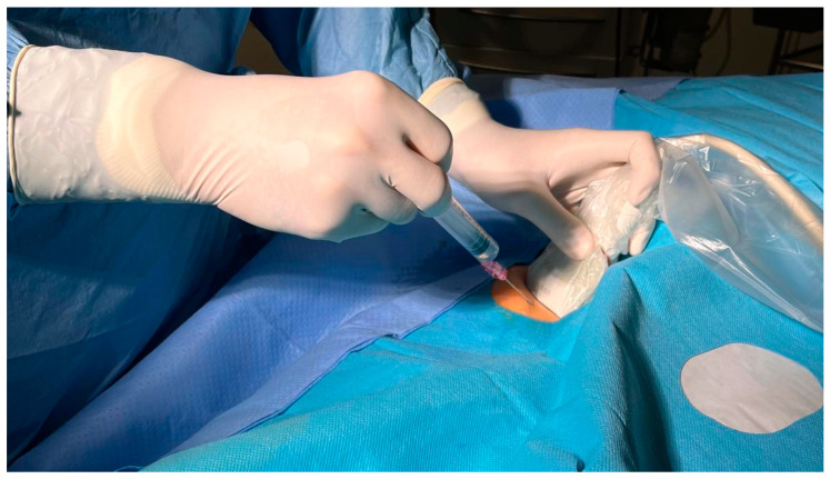
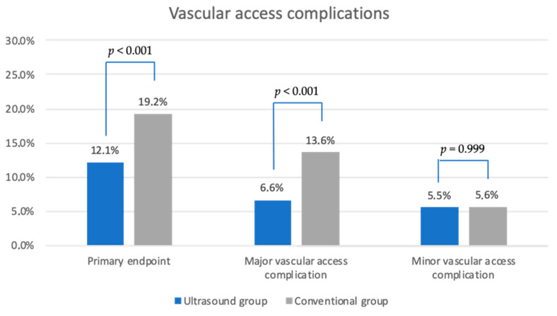
Background: In electrophysiological procedures, multiple punctures on the femoral vein can be necessary depending on the number of catheters required. The femoral vein is typically located indirectly by using its anatomical relationship to the artery as a reference. However, this conventional approach can lead to significant complications, including bleeding, peri-interventional transfusion, pseudoaneurysms, or arteriovenous fistulas. Despite these risks, there is limited evidence comparing the safety of ultrasound-guided venipuncture versus the conventional technique in electrophysiological procedures. Objective: This study aimed to evaluate the impact of ultrasound-guided venipuncture on vascular access complications in electrophysiological procedures and to identify associated risk factors. Methods: In this single-center trial, patients scheduled for electrophysiological procedures at Ulm University Heart Center, Germany, were enrolled between November 2021 and October 2023. Venipuncture in the groin was performed using either the conventional or an ultrasound-guided approach. The primary composite endpoint was defined as peri-interventional major vascular access complications (Bleeding Academic Research Consortium (BARC) ≥2 bleeding, pseudoaneurysms, arteriovenous fistulas, and peri-interventional transfusion) and minor complications (BARC 1). Results: A total of 1370 patients were included: 749 in the conventional group and 621 in the ultrasound group. The primary endpoint was achieved in 19.2% of the conventional group and 12.1% of the ultrasound group (p < 0.001). An increased sheath diameter and a higher number of venous accesses were identified as risk factors for the primary endpoint. Conclusions: Ultrasound guidance for venous groin puncture in electrophysiological procedures reduces access-related complications, supporting its use with careful attention to sheath size and number.
Keywords: arteriovenous fistula; bleeding; cardiology; electrophysiology; pseudoaneurysm; ultrasound; vascular access complication; venipuncture.
Conflict of interest statement
The authors declare no conflict of interest.
Figures


References
-
- Kupó P., Pap R., Sághy L., Tényi D., Bálint A., Debreceni D., Basu-Ray I., Komócsi A. Ultrasound Guidance for Femoral Venous Access in Electrophysiology Procedures—Systematic Review and Meta-Analysis. J. Interv. Card. Electrophysiol. 2020;59:407–414. doi: 10.1007/s10840-019-00683-z. - DOI - PMC - PubMed
-
- Cappato R., Calkins H., Chen S.-A., Davies W., Iesaka Y., Kalman J., Kim Y.-H., Klein G., Natale A., Packer D., et al. Updated Worldwide Survey on the Methods, Efficacy, and Safety of Catheter Ablation for Human Atrial Fibrillation. Circ. Arrhythm. Electrophysiol. 2010;3:32–38. doi: 10.1161/CIRCEP.109.859116. - DOI - PubMed
LinkOut - more resources
Full Text Sources
