. 2024 Oct 16;14(20):2980.
doi: 10.3390/ani14202980.

Novel Porcine Getah Virus from Diarrheal Piglets in Jiangxi Province, China: Prevalence, Genome Sequence, and Pathogenicity

Affiliations

Novel Porcine Getah Virus from Diarrheal Piglets in Jiangxi Province, China: Prevalence, Genome Sequence, and Pathogenicity

Jianhui Lan et al. Animals (Basel). .

Abstract

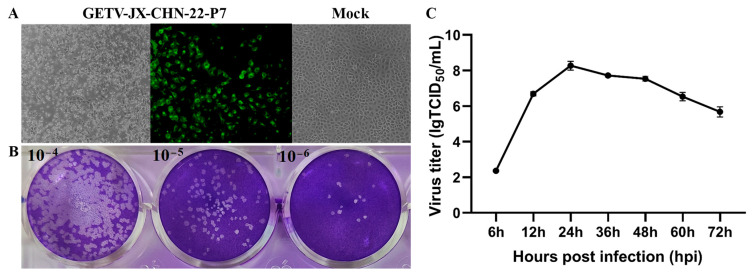
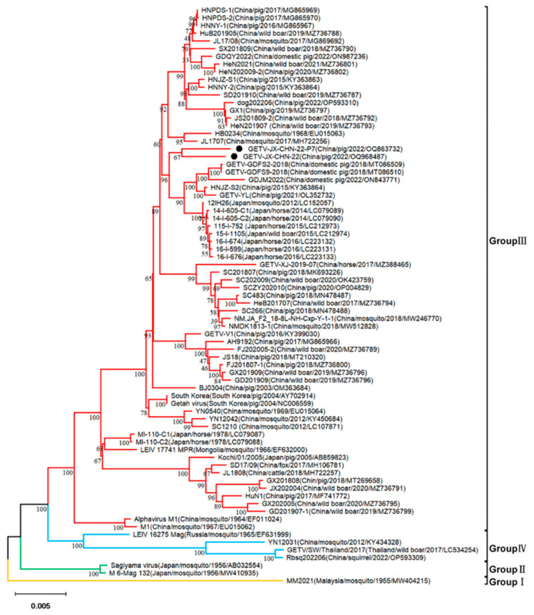
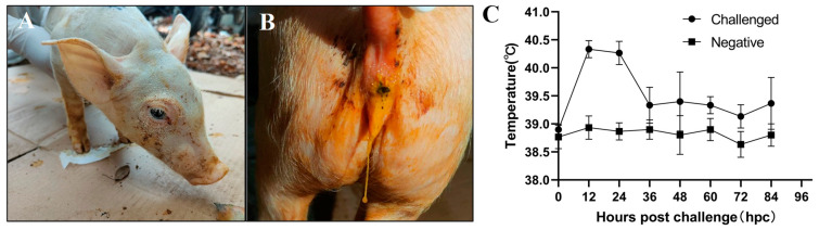
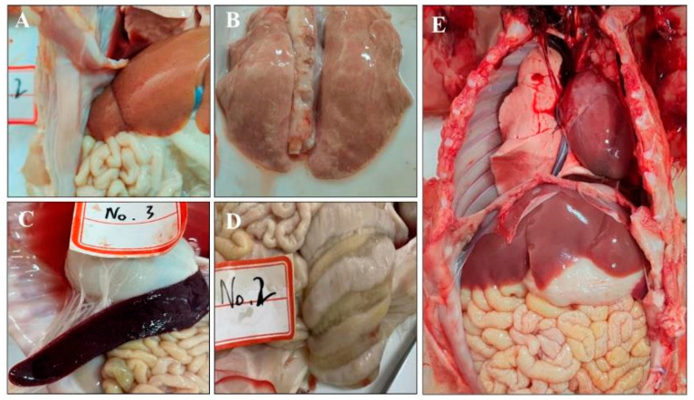
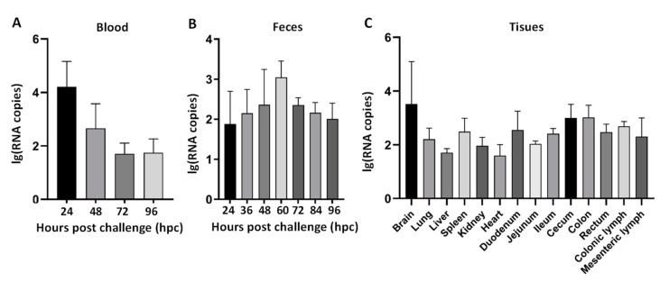
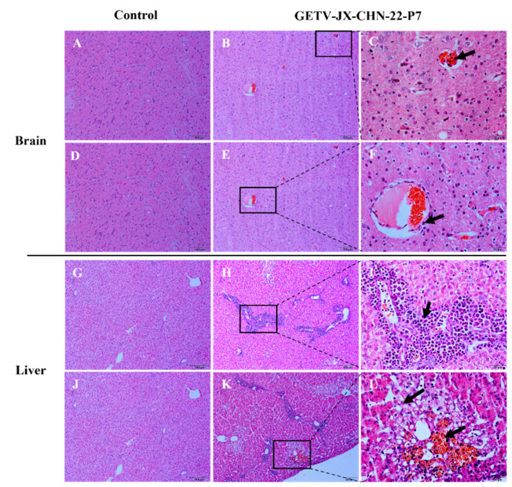
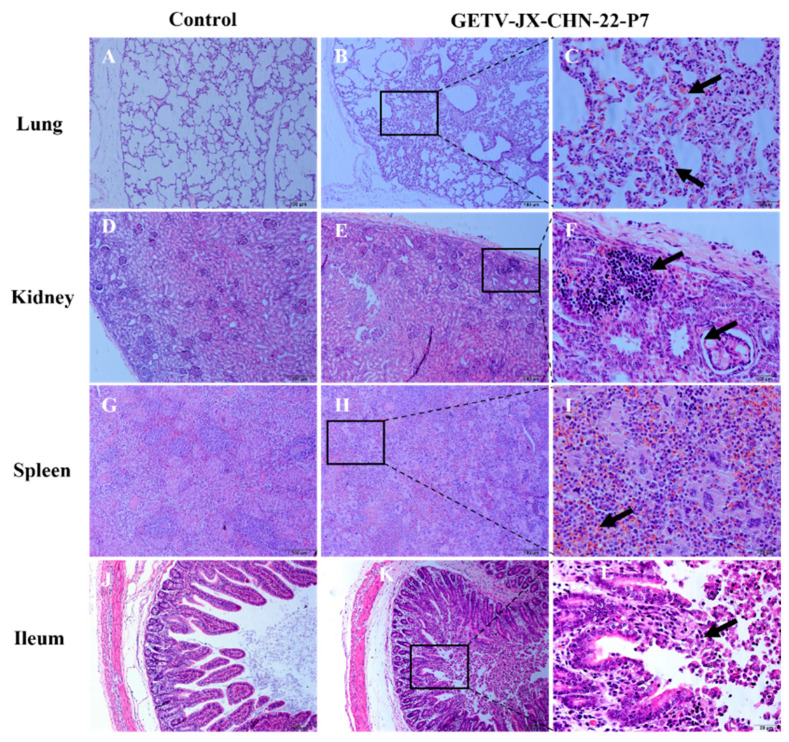
Getah virus (GETV) is a mosquito-borne virus belonging to the genus Alphavirus in the family Togaviridae. Its infection poses an increasing threat to animals and public health in China. In this study, an epidemiological survey of GETV on 46 pig farms in Jiangxi Province, China, was performed; GETV isolation and characterization were carried out, including a complete sequence determination and phylogenetic analysis; and pathogenicity of the GETV was experimentally investigated by inoculating newborn piglets with the isolated GETV strain. Epidemiological studies conducted on the organs of infected pigs, aborted piglets, and the blood of aborted sows sampled from pig farms in Jiangxi Province, China, demonstrated that 44 out of the 46 pig farms were positive for GETV, which is a positivity rate of 95.65% (44/46). Of the 411 samples tested, 47.93% (197/411) were found positive for GETV. A GETV strain called GETV-JX-CHN-22 was obtained, which showed stable proliferation in Vero cells. One-step growth curve results showed that the GETV-JX-CHN-22-P7 (passage 7) isolate reached a peak titer of 108.3 TCID50/mL at 24 hpi. An analysis of the whole-genome sequencing results showed that GETV-JX-CHN-22 (prototype) and GETV-JX-CHN-22-P7 shared nucleotide sequence similarities of 95.3% to 99.6% with 73 reference strains of GETV in GenBank. Genetic evolution analysis revealed that GETV-JX-CHN-22 and GETV-JX-CHN-22-P7 belonged to the GIII group, the same group members of most strains were reported in China. Animal inoculation experiments indicated that piglets exhibited typical symptoms and pathological changes of GETV infection after 24 h inoculation, which reproduced the pathogenicity of GETV field strain infections in piglets. To our knowledge, this study is the first report on the detection and isolation of porcine GETV associated with diarrhea from pig farms in Jiangxi Province, China. It is of great importance to study the infection spectrum, transmission mechanism, and public health significance of GETV. The results provide foundations for the genomic and biological (pathogenic) characteristics of the circulating GETV in Jiangxi Province, China.

Keywords: Getah virus; epidemiology; genomic sequencing; pathogenicity; swine; virus isolation.


Conflict of interest statement

The authors declare that the research was conducted in the absence of any commercial or financial relationships that could be construed as a potential conflict of interest.

Figures

Figure 1
Figure 1
Biological characteristics of GETV-JX-CHN-22-P7. (A) Cytopathic effects (CPEs) in Vero-81 cells (100× magnification) at 36 hpi observed in a microscope and IFA. (B) Plaques formed in Vero-81 cells after inoculation with 10−4, 10−5, and 10−6 dilutions of GETV-JX-CHN-22-P7. (C) Growth situation of GETV-JX-CHN-22-P7 at a multiplicity of infection (MOI) of 0.01 on Vero-81 cells. Viral titers were determined as TCID50, and all values are presented as mean ± SD from three independent experiments.
Figure 2
Figure 2
Phylogenetic analyses of complete genome (11,210 nt from ORF1 start codon to ORF2 stop codon, excluding 5′ and 3′ untranslated regions) of GETV. The phylogenetic tree was constructed using the p-distance-based neighbor-joining method with the MEGA v11 software. Bootstrap values (based on 1000 replicates) for each node are provided if they are >60%. The strain isolated in this study is labeled by a circle ●.
Figure 3
Figure 3
Clinical evaluation of piglets challenged with GETV strain GETV-JX-CHN-22-P7. (A) lethargy, (B) diarrhea, and (C) body temperature changes.
Figure 4
Figure 4
Gross lesions in piglets. Lesions in challenged piglets displayed petechial hemorrhages in the liver (A), pulmonary atrophy (B), splenic enlargement with hemorrhages (C), and colonic edema (D). The necropsies of the control group (E) revealed no significant abnormalities.
Figure 5
Figure 5
Virus load in blood (A), viral shedding in feces (B), and virus distribution in tissues at 96 hpi (C) of piglets challenged with GETV-JX-CHN-22-P7.
Figure 6
Figure 6
Clinical assessment of piglets challenged with GETV strain GETV-JX-CHN-22-P7. (A,D) Histologic view of brain tissues in control piglets. The scale bar indicates 100 µm. (B,E) Brain tissues showing congestion. (C,F) Higher magnification view of lesions boxed in panels B and E showing inflammatory cell infiltration in a vessel (arrows) and necrosis, along with mild neuronal degeneration. (G,J) Histopathologic examination of the brain and liver of control piglets revealed extensive inflammatory cell infiltration around the central veins, hepatocellular degeneration and necrosis with ballooning degeneration, and capillary rupture with hemorrhage (H,I,K,L). The scale bar in panels (A,B,D,E,G,H,J,K) indicates 100 µm, while the scale bar in panels (C,F,I,L) indicates 20 µm.
Figure 7
Figure 7
Clinical assessment of piglets challenged with GETV strain GETV-JX-CHN-22-P7. Histologic views of the lung, kidney, spleen, and ileum from control piglets (A,D,G,J) and challenged piglets (B,E,H,K). The scale bar indicates 100 µm. (C,F,I,L) Magnification of pathological changes in lung, kidney, spleen, and ileum in infected piglets, respectively. The scale bar in panels (A,B,D,E,G,H,J,K) indicates 100 µm, while the scale bar in panels (C,F,I,L) indicates 20 µm.

Similar articles

Cited by

References

    1. Chen R., Mukhopadhyay S., Merits A., Bolling B., Nasar F., Coffey L.L., Powers A., Weaver S.C. ICTV Report Consortium ICTV Virus Taxonomy Profile: Togaviridae. J. Gen. Virol. 2018;99:761–762. doi: 10.1099/jgv.0.001072. - DOI - PubMed
    1. Strauss J.H., Strauss E.G. The alphaviruses: Gene expression, replication, and evolution. Microbiol. Rev. 1994;58:491–562. doi: 10.1128/mr.58.3.491-562.1994. - DOI - PMC - PubMed
    1. Kamada M., Ando Y., Fukunaga Y., Kumanomido T., Imagawa H., Wada R., Akiyama Y. Equine Getah virus infection: Isolation of the virus from racehorses during an enzootic in Japan. Am. J. Trop. Med. Hyg. 1980;29:984–988. doi: 10.4269/ajtmh.1980.29.984. - DOI - PubMed
    1. Li X.D., Qiu F.X., Yang H., Rao Y.N., Calisher C.H. Isolation of Getah virus from mosquitos collected on Hainan Island, China, and results of a serosurvey. Southeast Asian J. Trop. Med. Public Health. 1992;23:730–734. - PubMed
    1. Ksiazek T., Trosper J., Cross J., Basaca-Sevilla V. Isolation of Getah virus from Nueva Ecija Province, Republic of the Philippines. Trans. R. Soc. Trop. Med. Hyg. 1981;75:312–313. doi: 10.1016/0035-9203(81)90346-1. - DOI - PubMed

LinkOut - more resources