Review
. 2024 Oct 15;13(20):6148.
doi: 10.3390/jcm13206148.

Role of Liquid-Based Cytology in the Endoscopic Diagnosis of Pancreatic Ductal Adenocarcinoma

Affiliations
Review

Role of Liquid-Based Cytology in the Endoscopic Diagnosis of Pancreatic Ductal Adenocarcinoma

Koh Kitagawa et al. J Clin Med. .

Abstract

Recently, endoscopic ultrasound-guided tissue acquisition (EUS-TA) has been widely used to diagnose pancreatic ductal adenocarcinoma (PDAC). The histological examination of core tissues acquired using novel biopsy needles is the primary diagnostic approach for patients with PDAC. However, in patients with early-stage PDAC, such as Stages 0 and I, EUS-TA can be challenging, and its diagnostic accuracy may be limited. This presents a clinical dilemma: The earlier that clinicians attempt to accurately diagnose PDAC, the more difficult it becomes to do so using EUS-TA. Liquid-based cytology (LBC) is a technique for preparing pathological specimens from liquefied cytology specimens by placing the collected material in a special fixative preservative fluid. LBC offers advantages, such as specimen optimization with reduced blood interference, a high cell-collection rate, and the simplicity of the procedure in the endoscopy room. The use of LBC may improve diagnostic accuracy, particularly for early-stage PDAC. Therefore, we emphasize that cytology remains a valuable tool for the endoscopic diagnosis of PDAC. In this review, we discuss the role of LBC in the endoscopic diagnosis of PDAC.

Keywords: cytology; endoscopic retrograde cholangiopancreatography; endoscopic ultrasound-guided fine needle aspiration; endoscopic ultrasound-guided fine needle biopsy; endoscopic ultrasound-guided tissue acquisition; histology; liquid-based cytology; pancreatic ductal adenocarcinoma.


Conflict of interest statement

The authors declare no conflicts of interest.

Figures
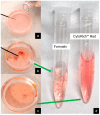
Figure 1
Figure 1
The specimen-handling flow in endoscopic ultrasound-guided tissue acquisition (EUS-TA) at our institution. (a) A small amount of dedicated preservative fluid (BD CytoRich Red Preservative; Becton Dickinson Japan, Tokyo, Japan) is placed in a Petri dish. (b) The specimen obtained by EUS-TA is pushed out of the puncture needle and promptly mixed directly with the preservative fluid. (c,d) Pieces of tissue with an apparent bulk are aspirated using a syringe, fixed in formalin, and prepared for histology. The residual paste or liquid specimens with preservative fluid are adapted to liquid-based cytology.
Figure 2
Figure 2
Laboratory protocol for processing samples from endoscopic ultrasound-guided tissue acquisition (EUS-TA) and endoscopic retrograde cholangiopancreatography (ERCP) at our institution.
Figure 3
Figure 3
Dedicated instruments for liquid-based cytology (SurePath method). (a) Dedicated glass slide (BD SurePath PreCoat slides; Becton Dickinson, Japan), with a charged center marked with a “+” (blue circle). (b) Settling chamber. (c) Tray for glass slides. (d) Dedicated glass slide and chamber mounted on trays.
Figure 4
Figure 4
Findings of Stage I (T1) pancreatic ductal adenocarcinoma. (a) Contrast-enhanced computed tomography (CE-CT) image. A pancreatic duct stenosis was observed in the tail of the pancreas, with dilation of the caudal pancreatic duct. Focal atrophy of the pancreatic parenchyma was noted at the site of the pancreatic duct stenosis, but no mass was visible on this CT. (b,c) Endoscopic ultrasound (EUS) images. A small hypoechoic mass less than 10 mm in diameter was identified at the site of the pancreatic duct stenosis (orange arrowheads). EUS-guided tissue acquisition was performed with a conventional 25-Gauge needle for fine needle aspiration. (d) Images of liquid-based cytology of specimens obtained from EUS-TA (Papanicolaou staining). The background of inflammatory cells and artifacts has been removed, revealing solitary, scattered tumor cells that can be evaluated. The cytological diagnosis was positive for adenocarcinoma. (e) Distal pancreatectomy was performed. Histopathologic findings revealed a 10 mm invasive carcinoma localized within the pancreas (hematoxylin–eosin staining).
Figure 5
Figure 5
Findings of Stage 0 pancreatic ductal adenocarcinoma. (a) Image of magnetic resonance cholangiopancreatography. Pancreatic duct stenosis was found in the head of the pancreas (orange arrowheads), and the caudal pancreatic duct was dilated. (b) Contrast-enhanced computed tomography (CE-CT) findings. There was pancreatic duct stenosis in the head of the pancreas (orange arrowheads), and the caudal pancreatic duct was dilated. However, no mass was visible on this CT scan. (c) Endoscopic ultrasound (EUS) image. No mass was visible at the site of pancreatic duct stenosis, even on EUS (orange arrowheads). (d) Endoscopic retrograde cholangiopancreatography. Pancreatography revealed stenosis of the main pancreatic duct in the head of the pancreas (orange arrowheads). An endoscopic nasopancreatic drainage tube was placed, and serial pancreatic juice aspiration cytology was performed. (e) Images of liquid-based cytology obtained via pancreatic juice cytology (Papanicolaou staining). The background of inflammatory cells and artifacts is removed, and solitary, scattered tumor cells can be evaluated. The cytological diagnosis was positive for adenocarcinoma. (f) A pancreatoduodenectomy was performed. Histopathologic findings on the resected specimen showed atypical columnar epithelial cells with chromatin-enriched enlarged nuclei proliferating in a hypopapillary fashion into the lumen of the pancreatic duct (hematoxylin–eosin staining). There was no obvious stromal invasion, and the diagnosis of carcinoma in situ was made.

Similar articles

References

    1. Itonaga M., Yasukawa S., Fukutake N., Ogura T., Asada M., Shimokawa T., Inatomi O., Nakai Y., Shiomi H., Nebiki H., et al. Comparison of 22-gauge Standard and Franseen Needles in EUS-guided Tissue Acquisition for Diagnosing Solid Pancreatic Lesions: A Multicenter Randomized Controlled Trial. Gastrointest. Endosc. 2022;96:57–66.e2. doi: 10.1016/j.gie.2022.02.005. - DOI - PubMed
    1. Kandel P., Nassar A., Gomez V., Raimondo M., Woodward T.A., Crook J.E., Fares N.S., Wallace M.B. Comparison of Endoscopic Ultrasound-guided Fine-needle Biopsy Versus Fine-needle Aspiration for Genomic Profiling and DNA Yield in Pancreatic Cancer: A Randomized Crossover Trial. Endoscopy. 2021;53:376–382. doi: 10.1055/a-1223-2171. - DOI - PubMed
    1. Hisada Y., Hijioka S., Ikeda G., Maehara K., Hashimoto T., Kitamura H., Harai S., Yoshinari M., Kawasaki Y., Murashima Y., et al. Proportion of Unresectable Pancreatic Cancer Specimens Obtained by Endoscopic Ultrasound-guided Tissue Acquisition Meeting the Oncoguide™ NCC Oncopanel System Analysis Suitability Criteria: A Single-arm, Phase II Clinical Trial. J. Gastroenterol. 2022;57:990–998. doi: 10.1007/s00535-022-01926-z. - DOI - PubMed
    1. Kawamura R., Ishii Y., Serikawa M., Tsuboi T., Tsushima K., Nakamura S., Hirano T., Ikemoto J., Kiyoshita Y., Saeki S., et al. Optimal Indication of Endoscopic Retrograde Pancreatography-based Cytology in the Preoperative Pathological Diagnosis of Pancreatic Ductal Adenocarcinoma. Pancreatology. 2022;22:414–420. doi: 10.1016/j.pan.2022.02.001. - DOI - PubMed
    1. Crinò S.F., Conti Bellocchi M.C., Bernardoni L., Manfrin E., Parisi A., Amodio A., De Pretis N., Frulloni L., Gabbrielli A. Diagnostic Yield of EUS-FNA of Small (≤15 mm) Solid Pancreatic Lesions Using a 25-gauge Needle. Hepatobiliary Pancreat. Dis. Int. 2018;17:70–74. doi: 10.1016/j.hbpd.2018.01.010. - DOI - PubMed

LinkOut - more resources