Case Reports
. 2024 Oct 12;12(10):2054.
doi: 10.3390/microorganisms12102054.

Capnocytophaga canimorsus Endocarditis Presenting with Leukocytoclastic Vasculitis and Glomerulonephritis

Affiliations
Case Reports

Capnocytophaga canimorsus Endocarditis Presenting with Leukocytoclastic Vasculitis and Glomerulonephritis

Divya Chandramohan et al. Microorganisms. .

Abstract

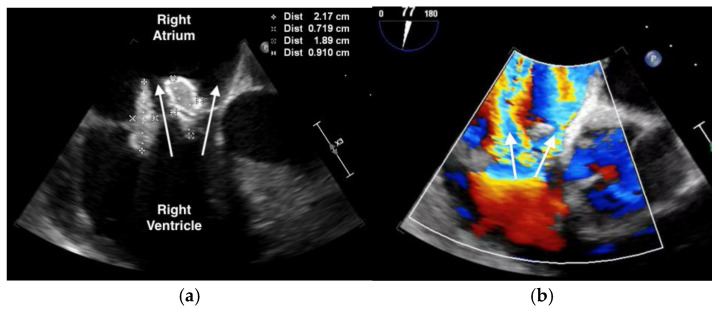
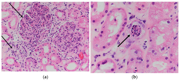
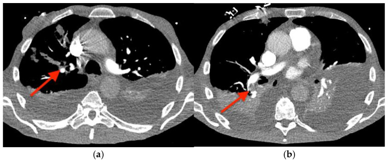
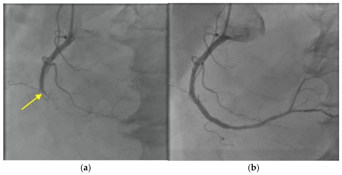
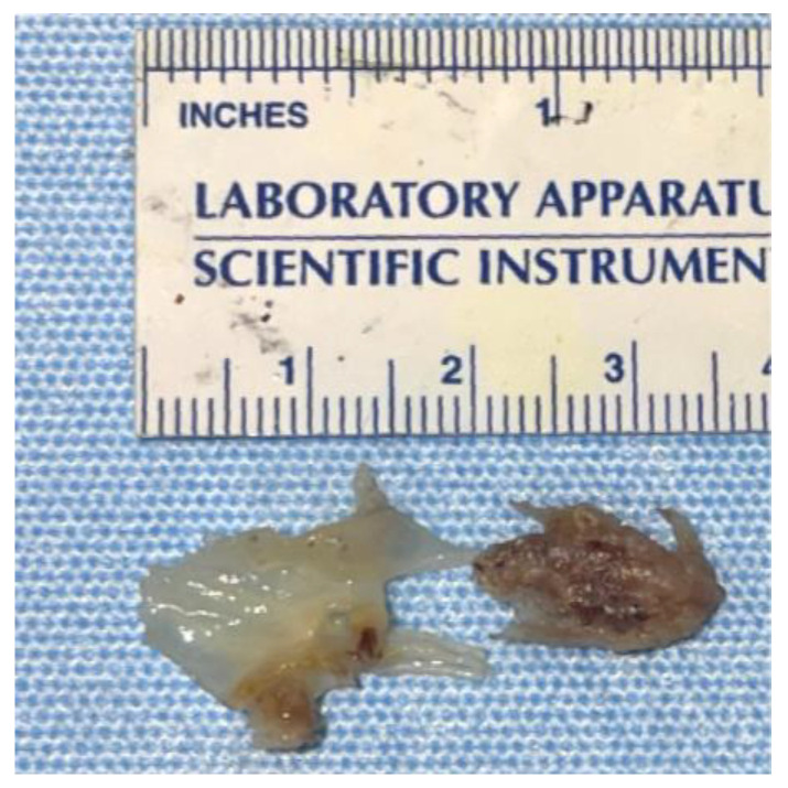
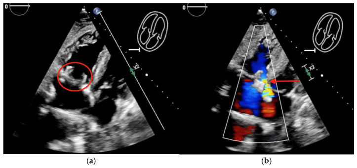
Capnocytophaga canimorsus is a gram-negative bacterium commonly found in the saliva of dogs and cats. Despite the frequency of animal bites, infection with Capnocytophaga species is rare, and severe infections are usually associated with underlying risk factors, such as alcohol use disorder, asplenia, or immunosuppression. We describe a case of a man who presented with a purpuric rash, lower extremity edema, and acute renal failure and was found to have tricuspid valve endocarditis and infection-associated glomerulonephritis due to C. canimorsus. Despite treatment with cefepime, the vegetation increased in size and valvular function worsened. He was readmitted with an inferior wall myocardial infarction, heart failure, and pulmonary embolism. He underwent an urgent tricuspid valve replacement with a bioprosthetic valve. A 16S ribosomal RNA amplicon sequencing performed on the resected valve tissue verified involvement of C. canimorsus. Post-operatively, he had several episodes of gastrointestinal hemorrhage requiring multiple endoscopic interventions and arterial embolization. The recurrent gastrointestinal hemorrhage combined with his severe functional decline ultimately led to his death. This patient had an uncommon presentation with leukocytoclastic vasculitis and infection-associated glomerulonephritis, which revealed an underlying diagnosis of infective endocarditis due to C. canimorsus, a rare gram-negative bacterial etiology of infective endocarditis.

Keywords: Capnocytophaga; dog bite; endocarditis; glomerulonephritis; tricuspid valve; vasculitis.


Conflict of interest statement

The authors declare no conflicts of interest.

Figures

Figure 1
Figure 1
Rash associated with C. canimorsus bacteremia and endocarditis. Purpuric rash of (a) the right palm and (b) the right lower extremity. The left lower extremity was similar, but the rash was less pronounced.
Figure 2
Figure 2
Transesophageal echocardiogram showing: (a) large, multi-lobed tricuspid valve vegetations associated with valve perforation and (b) severe tricuspid valve regurgitation (white arrow).
Figure 3
Figure 3
Characteristic pinpoint colonies of C. canimorsus, as seen on blood agar.
Figure 4
Figure 4
Renal biopsy showing diffuse acute proliferative glomerulonephritis: (a) global glomerular enlargement and hypercellularity with dense neutrophilic and histiocytic infiltrate, as well as interstitial acute inflammation and edema (black arrow) (Hematoxylin and Eosin (H&E), 200× magnification) and (b) endothelial cell swelling with capillary luminal obstruction by acute inflammatory cells (black arrow) (H&E, 400× magnification).
Figure 5
Figure 5
Computed tomography angiogram of the chest showing pulmonary emboli (red arrows) in: (a) the right upper lobe and (b) the right lower lobe.
Figure 6
Figure 6
Cardiac catheterization with right coronary artery angiogram showing: (a) total occlusion of the right coronary artery (yellow arrow), followed by (b) restoration of blood flow after deployment of a drug-eluting stent.
Figure 7
Figure 7
Resected tricuspid valve tissue showing the attached vegetation.
Figure 8
Figure 8
Postoperative transthoracic echocardiogram showing: (a) new bioprosthetic tricuspid valve (red circle) and (b) residual paravalvular leak (red arrow).

References

    1. Lion C., Escande F., Burdin J.C. Capnocytophaga canimorsus infections in human: Review of the literature and cases report. Eur. J. Epidemiol. 1996;12:521–533. doi: 10.1007/BF00144007. - DOI - PubMed
    1. Hino C., Veltman J. Capnocytophaga tricuspid valve endocarditis: A case report and literature review. Access Microbiol. 2022;4:acmi000355. doi: 10.1099/acmi.0.000355. - DOI - PMC - PubMed
    1. Archer S.L. Dysgonic fermenter 2 infection resulting in chronic glomerulonephritis. Can. Med. Assoc. J. 1985;132:657–660. - PMC - PubMed
    1. Kannan S., Mattoo T.K. Diffuse crescentic glomerulonephritis in bacterial endocarditis. Pediatr. Nephrol. 2001;16:423–428. doi: 10.1007/s004670000550. - DOI - PubMed
    1. Li M., Kim J.B., Sastry B.K.S., Chen M. Infective endocarditis. Lancet. 2024;404:377–392. doi: 10.1016/S0140-6736(24)01098-5. - DOI - PubMed

Publication types

LinkOut - more resources