. 2024 Sep 28;16(10):1267.
doi: 10.3390/pharmaceutics16101267.

A Modified Cell-Penetrating Peptide Enhances Insulin and Oxytocin Delivery across an RPMI 2650 Nasal Epithelial Cell Barrier In Vitro

Affiliations

A Modified Cell-Penetrating Peptide Enhances Insulin and Oxytocin Delivery across an RPMI 2650 Nasal Epithelial Cell Barrier In Vitro

Sara Wong et al. Pharmaceutics. .

Abstract

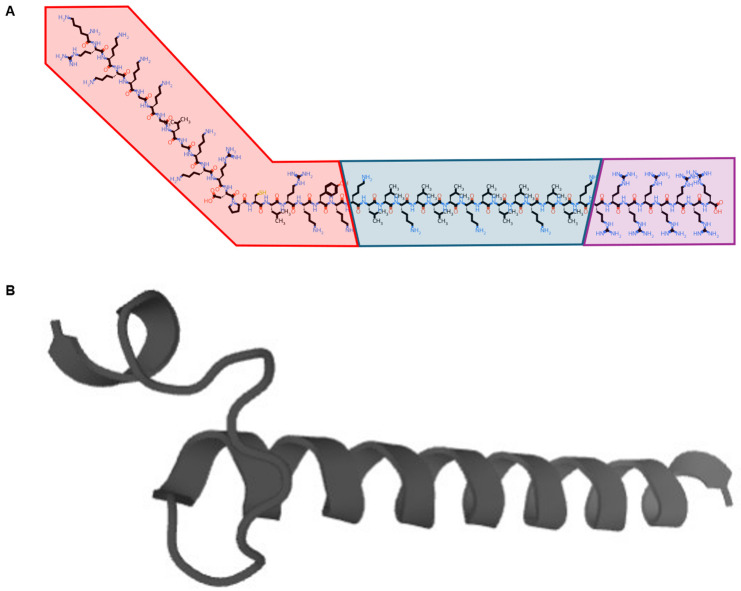
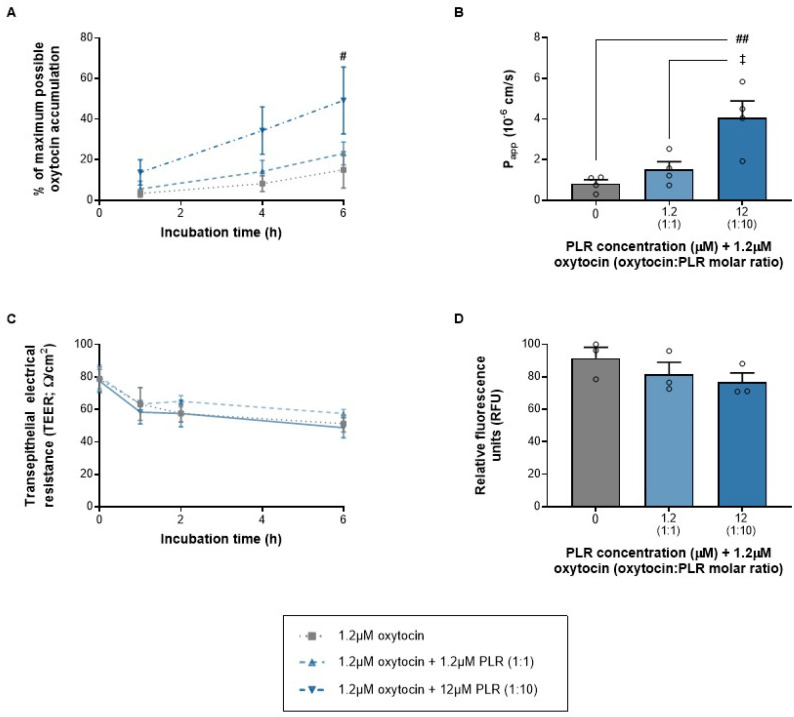
Background/Objectives: Peptide-based treatments represent an expanding area and require innovative approaches to enhance bioavailability. Combination with cell-penetrating peptides (CPPs) is an attractive strategy to improve non-invasive delivery across nasal epithelial barriers for systemic and direct nose-to-brain transport. We previously developed a modified CPP system termed Glycosaminoglycan-binding Enhanced Transduction (GET) that improves insulin delivery across gastrointestinal epithelium. It contains a membrane docking sequence to promote cellular interactions (P21), a cationic polyarginine domain to stimulate uptake (8R) and an endosomal escaping sequence to maximize availability for onward distribution (LK15). It is synthesized as a single 44-residue peptide (P21-LK15-8R; PLR).

Methods: The current research used in vitro assays for a novel exploration of PLR's ability to improve the transport of two contrasting peptides, insulin (51 residues, net negative charge) and oxytocin (9 residues, weak positive charge) across an RPMI 2650 human nasal epithelial cell barrier cultured at the air-liquid interface.

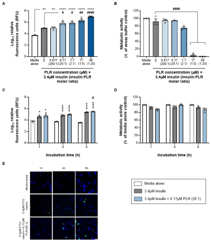
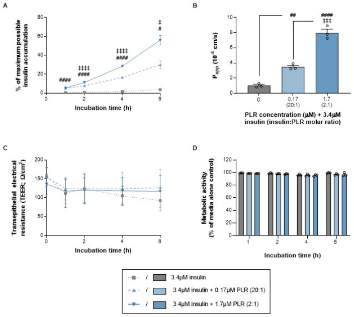
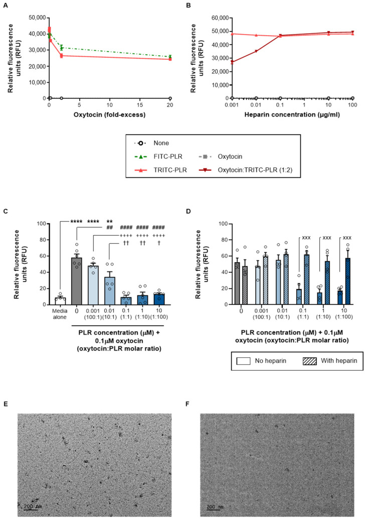
Results: PLR enhanced insulin transcytosis over a 6 h period by 7.8-fold when used at a 2:1 molar ratio of insulin/PLR (p < 0.0001 versus insulin alone). Enhanced oxytocin transcytosis (5-fold) occurred with a 1:10 ratio of oytocin/PLR (p < 0.01). Importantly, these were independent of any impact on transepithelial electrical resistance (TEER) or cell viability (p > 0.05).

Conclusions: We advocate the continued evaluation of insulin-PLR and oxytocin-PLR formulations, including longer-term assessments of ciliotoxicity and cytotoxicity in vitro followed by in vivo assessments of systemic and nose-to-brain delivery.

Keywords: RPMI 2650; cell-penetrating peptide; glycosaminoglycan-GAG-binding enhanced transduction; insulin; nasal drug delivery; oxytocin; transcytosis; transepithelial delivery.


Conflict of interest statement

The authors declare no conflicts of interest.

Figures

Figure 1
Figure 1
The amino acid sequence of the PLR peptide is KRKKKGKGLGKKRDPCLRKYKKLLKLLLKLLLKLLKRRRRRRRR. The images show (A) a simplified molecular-input line-entry system (SMILES) notation, with the heparan-binding domain from heparin-binding epidermal growth factor (P21, red), endosomal escaping sequence (LK15, blue) and cationic polyarginine CPP (8R, purple; produced using the PepSMI tool by NovoPro, https://www.novoprolabs.com/tools/convert-peptide-to-smiles-string, accessed on 22 September 2024) and (B) a two-dimensional model (produced using the PEP-FOLD3 tool [11] https://bioserv.rpbs.univ-paris-diderot.fr/services/PEP-FOLD3/, accessed on 22 September 2024).
Figure 2
Figure 2
PLR increases insulin uptake into RPMI 2650 nasal epithelial cells at concentrations that do not reduce cell viability. Data are shown as mean ± s.e.m. (with individual data points also shown) for (A) insulin uptake and (B) cell viability following 24 h of incubation with insulin alone (gray bars) and increasing PLR concentrations (blue bars), (C) insulin uptake and (D) cell viability following 1–6 h of incubation with insulin alone (gray bars) or 20:1 molar ratio of insulin/PLR (blue bars), as well as (E) representative images showing cells that have taken up FITC-insulin (green) as proportion of total cells, labeled with DAPI (blue); scale bar = 25 μm. */**/**** p < 0.05/0.01/0.0001 versus media alone (control) and #/##/#### p < 0.05/0.01/0.0001 versus insulin alone (one-way ANOVA with Tukey’s multiple-comparison post hoc test or two-way ANOVA with Sidak’s multiple-comparison post hoc test). Viability data from (media alone) control (B,D) have zero variance and were excluded from statistical analyses.
Figure 3
Figure 3
PLR increases insulin transcytosis across an RPMI 2650 nasal epithelial cell barrier (cultured on transwell inserts at the air–liquid interface) at concentrations that do not reduce transepithelial electrical resistance (TEER) or cell viability. The data are shown as the mean ± s.e.m. (with individual data points also shown on the histograms) for (A) the time course of insulin accumulation in the basal chamber; (B) apparent permeability (Papp) across the 6 h period as a whole; and (C) TEER and (D) cell viability time courses following incubation with insulin alone (gray lines/bars) and increasing PLR concentrations (blue lines/bars). #/##/#### p < 0.05/0.01/0.0001 for both 20:1 and 2:1 molar ratios of insulin/PLR versus insulin alone and ‡/‡‡‡/‡‡‡‡ p < 0.05/0.001/0.0001 for a 2:1 molar ratio of insulin/PLR versus 20:1 (one-way ANOVA, two-way ANOVA or two-way repeated-measures ANOVA with Geisser–Greenhouse’s correction for unequal variance, all followed by Tukey’s multiple-comparison post hoc test).
Figure 4
Figure 4
PLR complexes with oxytocin in a reversible, heparin-sensitive manner. The data are shown as the mean ± s.e.m. (with individual data points also shown on the histograms) for (A) the quenching of FITC-PLR (green dotted line) and TRITC-PLR (red solid line) fluorescence by increasing concentrations of oxytocin; (B) the dequenching of oxytocin/TRITC-PLR fluorescence by increasing concentrations of heparin (dark red line); as well as (C) the bioactivity of oxytocin alone (gray bars) in Hs 578T cells, which is reduced by increasing PLR concentrations (blue bars) and (D) restored by heparin (diagonal shaded bars). The images were obtained from a TEM examination of (E) 1:1 and (F) 1:10 ratios of oxytocin/PLR. **/**** p < 0.01/0.0001 versus media alone (control), ##/#### p < 0.01/0.0001 versus oxytocin alone, ++++ p < 0.001 versus a 100:1 molar ratio of oxytocin/PLR, †/†† p < 0.05/0.01 versus a 10:1 molar ratio of oxytocin/PLR, and xxx p < 0.001 versus the same oxytocin/PLR ratio in the absence of heparin (one-way ANOVA or two-way ANOVA with Tukey’s multiple-comparison post hoc test).
Figure 5
Figure 5
PLR increases oxytocin transcytosis across an RPMI 2650 nasal epithelial cell barrier (cultured on transwell inserts at the air–liquid interface) at concentrations that do not reduce the transepithelial electrical resistance (TEER) or cell viability. The data are shown as the mean ± s.e.m. (with individual data points also shown on the histograms) for (A) the time course of oxytocin accumulation in the basal chamber; (B) apparent permeability (Papp) across the 6 h period as a whole; as well as (C) the TEER across the time course of transcytosis measurements and (D) cell viability at the end of the 6 h period following incubation with oxytocin alone (gray lines/bars) and increasing PLR concentrations (blue lines/bars). #/## p < 0.05/0.01 for both the 1:10 molar oxytocin/PLR ratio versus oxytocin alone and p < 0.05 for the 1:10 molar oxytocin/PLR ratio versus 1:1 (one-way ANOVA, two-way ANOVA or two-way repeated-measures ANOVA with Geisser–Greenhouse’s correction for unequal variance, all followed by Tukey’s multiple-comparison post hoc test).

References

    1. Rossino G., Marchese E., Galli G., Verde F., Finizio M., Serra M., Linciano P., Collina S. Peptides as therapeutic agents: Challenges and opportunities in the green transition era. Molecules. 2023;28:7165. doi: 10.3390/molecules28207165. - DOI - PMC - PubMed
    1. Kristensen M., Nielsen H.M. Cell-penetrating peptides as tools to enhance non-injectable delivery of biopharmaceuticals. Tissue Barriers. 2016;4:e1178369. doi: 10.1080/21688370.2016.1178369. - DOI - PMC - PubMed
    1. De Martini L.B., Sulmona C., Brambilla L., Rossi D. Cell-penetrating peptides as valuable tools for nose-to-brain delivery of biological drugs. Cells. 2023;12:1643. doi: 10.3390/cells12121643. - DOI - PMC - PubMed
    1. Luo D., Ni X., Yang H., Feng L., Chen Z., Bai L. A comprehensive review of advanced nasal delivery: Specially insulin and calcitonin. Eur. J. Pharm. Sci. 2024;192:106630. doi: 10.1016/j.ejps.2023.106630. - DOI - PubMed
    1. Dixon J.E., Osman G., Morris G.E., Markides H., Rotherham M., Bayoussef Z., El Haj A.J., Denning C., Shakesheff K.M. Highly efficient delivery of functional cargoes by the synergistic effect of GAG binding motifs and cell-penetrating peptides. Proc. Natl. Acad. Sci. USA. 2016;113:E291–E299. doi: 10.1073/pnas.1518634113. - DOI - PMC - PubMed

LinkOut - more resources