. 2024 Sep 30;16(10):1282.
doi: 10.3390/pharmaceutics16101282.

Synergistic Enhancement of Carboplatin Efficacy through pH-Sensitive Nanoparticles Formulated Using Naturally Derived Boswellia Extract for Colorectal Cancer Therapy

Affiliations

Synergistic Enhancement of Carboplatin Efficacy through pH-Sensitive Nanoparticles Formulated Using Naturally Derived Boswellia Extract for Colorectal Cancer Therapy

Sherif Ashraf Fahmy et al. Pharmaceutics. .

Abstract

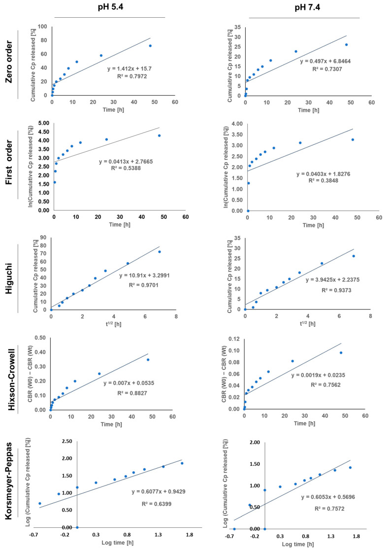
Carboplatin (Cp) is a potent chemotherapeutic agent, but its effectiveness is constrained by its associated side effects. Frankincense, an oleo-gum resin from the Boswellia sacra tree, has demonstrated cytotoxic activity against cancer cells. This study explored the synergistic potential of nanoparticles formulated from Boswellia sacra methanolic extract (BME), to enhance the therapeutic efficacy of Cp at reduced doses. Nanoparticles were prepared via the nanoprecipitation method, loaded with Cp, and coated with positively charged chitosan (CS) for enhanced cell interaction, yielding Cp@CS/BME NPs with an average size of 160.2 ± 4.6 nm and a zeta potential of 12.7 ± 1.5 mV. In vitro release studies revealed a pH-sensitive release profile, with higher release rates at pH 5.4 than at pH 7.4, highlighting the potential for targeted drug delivery in acidic tumor environments. In vitro studies on HT-29 and Caco-2 colorectal cancer cell lines demonstrated the nanoformulation's ability to significantly increase Cp uptake and cytotoxic activity. Apoptosis assays further confirmed increased induction of cell death with Cp@CS/BME NPs. Cell-cycle analysis revealed that treatment with Cp@CS/BME NPs led to a significant increase in the sub-G1 phase, indicative of enhanced apoptosis, and a marked decrease in the G1-phase population coupled with an increased G2/M-phase arrest in both cell lines. Further gene expression analysis demonstrated a substantial downregulation of the anti-apoptotic gene Bcl-2 and an upregulation of the pro-apoptotic genes Bax, PUMA, and BID following treatment with Cp@CS/BME NPs. Thus, this study presents a promising and innovative strategy for enhancing the therapeutic efficacy of chemotherapeutic agents using naturally derived ingredients while limiting the side effects.

Keywords: chemotherapy; colorectal cancer; nanomedicine; nanoprecipitation; pH-sensitive release.


Conflict of interest statement

The authors declare no conflicts of interest.

Figures

Figure 1
Figure 1
LC-ESI-MS/MS ion chromatogram (in the negative ion mode) of the Boswellia sacra oleo-gum resin methanolic extract.
Figure 2
Figure 2
Physicochemical characterization of Cp@CS/BME NPs: (A) TEM image illustrating the morphology of Cp@CS/BME NPs; scale bar = 200 nm. (B) Zeta potential of BME NPs and Cp@CS/BME NPs. (C) In vitro release profile of Cp from Cp@CS/BME NPs into acetate buffer (pH 5.4) and phosphate buffer (pH 7.4). Data are presented as the mean ± SD; n = 3.
Figure 3
Figure 3
The release kinetics of Cp from BME NPs at pH 5.4 and 7.4, were determined using mathematical kinetics models.
Figure 4
Figure 4
The uptake of Cp in its free or Cp@CS/BME NPs forms by HT-29 cells (left panel) and Caco-2 cells (right panel). Results represent the mean value ± SD (n = 3). The symbols *, **, *** indicate statistical significance from control (Cp) at p-values < 0.05, <0.01, and <0.001, respectively.
Figure 5
Figure 5
Dose–response curves showing the inhibitory effects of BME, BME NPs, Cp, and Cp@CS/BME NPs on the HT-29 cells (top panel) and caco-2 cells (bottom panel). Results represent the mean value ± SD (n = 3).
Figure 6
Figure 6
Apoptotic effects were observed in the HT-29 and Caco-2 cell lines following 48 h of exposure to Cp@CS/BME NPs. The top panel presents cytograms of Annexin V/propidium iodide-stained HT-29 cells, comparing untreated cells as the control group (A) with Cp@CS/BME NPs-treated cells (B). The corresponding bar graph (C) quantifies the percentages of necrotic, apoptotic, and viable HT-29 cells. The bottom panel depicts the cytograms for Annexin V/propidium iodide-stained Caco-2 cells, showing untreated control cells (D) and Cp@CS/BME NPs-treated cells (E). The bar graph (F) illustrates the proportions of necrotic, apoptotic, and viable Caco-2 cells. ** p < 0.01, *** p < 0.001, and **** p < 0.0001. Results represent the mean value ± SD (n = 3).
Figure 7
Figure 7
Cell-cycle analysis of HT-29 and Caco-2 cells treated with Cp@CS/BME NPs. The top panel presents cytograms of untreated (control) HT-29 cells (A) and HT-29 cells treated with Cp@CS/BME NPs (B), accompanied by a bar graph illustrating the cell-cycle distribution of HT-29 cells (C). The bottom panel shows the cytograms of untreated (control) Caco-2 cells (D) and Caco-2 cells treated with Cp@CS/BME NPs (E), along with a bar graph showing the cell-cycle distribution of Caco-2 cells (F). * p < 0.05, ** p < 0.01, *** p < 0.001, and **** p < 0.0001. Results represent the mean value ± SD (n = 3).
Figure 8
Figure 8
RT-qPCR analysis of target genes in HT-29 (top panel) and Caco-2 cells (bottom panel) following 48 h incubation with Cp or Cp@CS/BME NPs. Gene expression levels are normalized to β-actin and presented as the mean ± SD of three independent experiments; (#) and (*) indicate statistical significance compared to the control and Cp-treated groups, respectively. ** p < 0.01, ## p < 0.01, *** p < 0.001 and ### p < 0.001.

References

    1. Biller L.H., Schrag D. Diagnosis and Treatment of Metastatic Colorectal Cancer: A Review. JAMA. 2021;325:669–685. doi: 10.1001/jama.2021.0106. - DOI - PubMed
    1. Xi Y., Xu P. Global colorectal cancer burden in 2020 and projections to 2040. Transl. Oncol. 2021;14:101174. doi: 10.1016/j.tranon.2021.101174. - DOI - PMC - PubMed
    1. Buyana B., Naki T., Alven S., Aderibigbe B.A. Nanoparticles Loaded with Platinum Drugs for Colorectal Cancer Therapy. Int. J. Mol. Sci. 2022;23:11261. doi: 10.3390/ijms231911261. - DOI - PMC - PubMed
    1. Ritacco I., Al Assy M., Abd El-Rahman M.K., Fahmy S.A., Russo N., Shoeib T., Sicilia E. Hydrolysis in Acidic Environment and Degradation of Satraplatin: A Joint Experimental and Theoretical Investigation. Inorg. Chem. 2017;56:6013–6026. doi: 10.1021/acs.inorgchem.7b00945. - DOI - PubMed
    1. Sedky N.K., Abdel-Kader N.M., Issa M.Y., Abdelhady M.M.M., Shamma S.N., Bakowsky U., Fahmy S.A. Co-Delivery of Ylang Ylang Oil of Cananga odorata and Oxaliplatin Using Intelligent pH-Sensitive Lipid-Based Nanovesicles for the Effective Treatment of Triple-Negative Breast Cancer. Int. J. Mol. Sci. 2023;24:8392. doi: 10.3390/ijms24098392. - DOI - PMC - PubMed

LinkOut - more resources