. 2024 Oct 11;17(10):1358.
doi: 10.3390/ph17101358.

Vesicle-Transported Multidrug Resistance as a Possible Therapeutic Target of Natural Compounds

Affiliations

Vesicle-Transported Multidrug Resistance as a Possible Therapeutic Target of Natural Compounds

Salvatrice Rigogliuso et al. Pharmaceuticals (Basel). .

Abstract

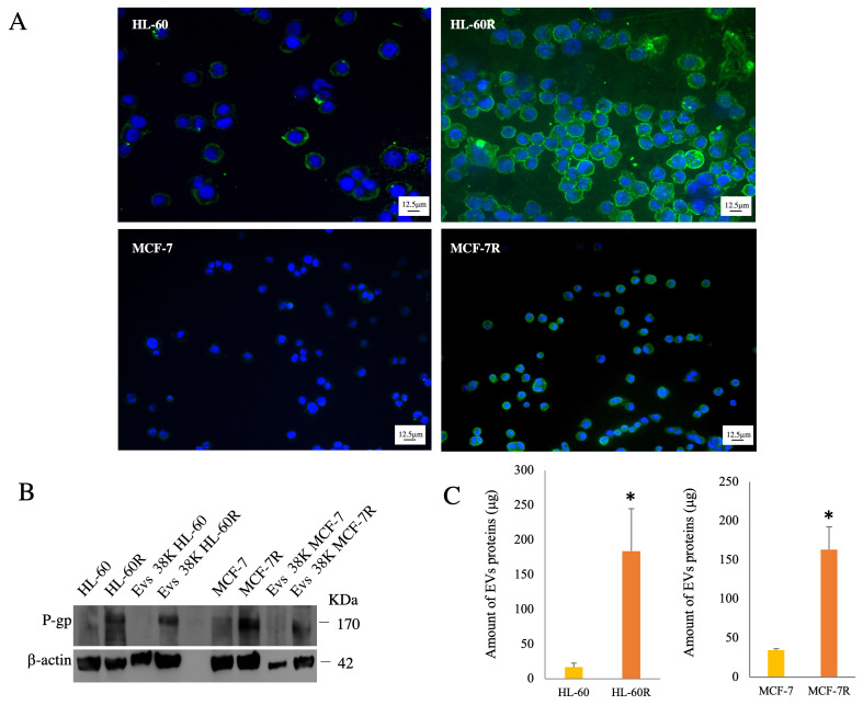
Background/objectives: A key role of extracellular vesicles (EVs) is mediating both cell-cell and cell-stroma communication in pathological/physiological conditions. EVs from resistant tumor cells can transport different molecules like P-glycoprotein (P-gp), acting as a shuttle between donor and recipient cells, resulting in a phenotypic change. The aim of our work was to isolate, characterize, and inhibit the release of EVs in two multidrug resistance (MDR) cancer models: MCF-7R (breast cancer cell line) and HL-60R (acute myeloid leukemia cell line).

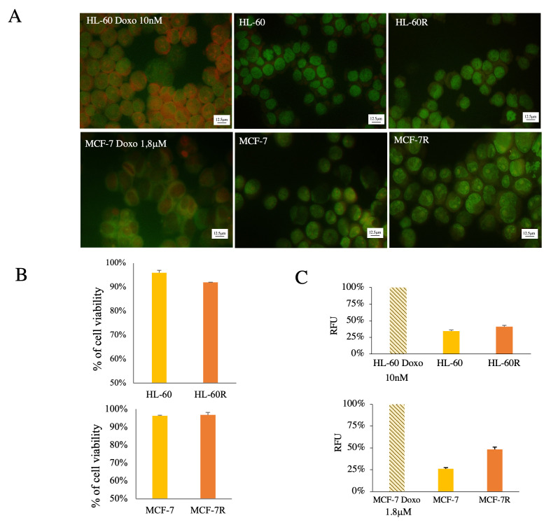
Methods: The existence of P-gp in EVs from MDR cells was confirmed by Western blotting assays. The characterization of EVs was carried out by evaluating the size using NTA and the presence of specific markers such as CD63, Hsp70 and Syntenin. The ability of HL-60R and MCF-7R to perform horizontal transfer of P-gp via EVs to sensitive cells was assessed using three different methods. The acquisition of resistance and its inhibition in recipient cells was confirmed by MTS 3-(4,5-dimethylthiazol-2-yl)-5-(3-carboxymethoxyphenyl)-2-(4-sulfophenyl)-2H-tetrazolium (MTS) assay.

Results: Our data showed that cell lines (MDR) release P-gp-loaded EVs, unlike sensitive cells. The acquisition of resistance determined by the incorporation of P-gp into the membrane of sensitive cells was confirmed by the reduced cytotoxic activity of doxorubicin. Natural compounds such as curcumin, lupeol, and heptacosane can block vesicular transfer and restore the sensitivity of HL-60 and MCF-7 cells.

Conclusions: Our study demonstrates that natural inhibitors able to reverse this mechanism may represent a new therapeutic strategy to limit the propagation of the resistant phenotype.

Keywords: P-glycoprotein; acute myeloid leukemia; breast cancer; cancer drug resistance; extracellular vesicles.


Conflict of interest statement

All authors declare there are no conflicts of interest.

Figures

Figure 1
Figure 1
Evaluation of P-gp expression in EVs isolated from resistant cell lines. (A) Immunofluorescence analysis of HL-60, HL-60R, MCF-7, and MCF-7R cells. Nuclei (DAPI Blue); P-gp (green). (B) Western blotting analysis of cell extract and 38,000 rpm fractions (38K) of EVs isolated, respectively, from HL-60R and MCF-7R cell lines. (C) Comparison of the amount of protein content in EVs released from both sensitive cell lines compared to resistant cell lines. Data were normalized to the same number of cells: HL-60R vs. HL-60 * p < 0.05; MCF-7R vs. MCF-7 * p < 0.01 (Tukey’s test).
Figure 2
Figure 2
Analysis of cells producing EVs (38K) with AO staining: (A) fluorescence microscopy images; (B) Trypan Blue exclusion assay; (C) spectrofluorometric analysis. Fluorescence emission of the cells was compared to positive control (apoptosis induced by doxorubicin treatment; RFU—Relative Fluorescence Units).
Figure 3
Figure 3
Characterization analysis of EVs isolated from resistant cell lines. (A) Western blot analysis of the EV markers, Hsp-70, CD63, and Syntenin. (B) Size distributions of EV fractions isolated, respectively, from HL-60R and MCF-7R cell lines measured by nanoparticle tracking analysis (NTA). (C) Separation of heterogeneous EV populations by differential ultracentrifugation and relative %; Western blotting analysis for P-gp and Syntenin expression in a 35k EV fraction.
Figure 4
Figure 4
Analysis of the horizontal transfer of resistance. Representative image of evaluation of P-gp expression after following treatment of the sensitive HL-60 and MCF-7 cell lines: EVs isolated from the respective resistant cell lines, with the medium conditioned by the cells of the respective resistant lines or by the co-culture growth of the sensitive and resistant line in a Transwell plate. Nuclei (DAPI Blue); P-gp (green); magnification 20× and scale bar was 25 µm.
Figure 5
Figure 5
(A) Cells were exposed to Doxorubicin (1.8 μM) for 16 h after co-culture. Cell viability was assessed by an MTS assay. Letters indicate significant differences (Tukey’s test) in cell viability among the concentrations of each cell line (Cytochalasin, Curcumin, and Heptacosane vs. co-culture + Doxorubicin p < 0.005; Cytochalasin vs. Lupeol p < 0.001; Curcumin vs. Lupeol and Heptacosane p < 0.05). Treatments are likened to the control: * p < 0.005; ** p < 0.001. (B) Cells were exposed to Doxorubicin (9.2 μM) for 24 h after co-culture. Cell viability was assessed by cell counting. Letters indicate significant differences (Tukey test) in cell viability among the concentrations (Cytochalasin and Heptacosane vs. co-culture + Doxorubicin p < 0.05; Cytochalasin vs. Lupeol and Curcumin p < 0.05). Treatments are likened to the control: * p < 0.05.
Figure 6
Figure 6
Co-culture of MCF-7 and MCF-7R cell spheroids. MCF-7 and MCF-7R cell spheroids in co-culture for 24 h, labeled with DAPI (blue nuclei) and anti-P-gp mAb (green fluorescence). (A) Immunofluorescence at time zero and after 24 h of co-culture with an anti-P-gp antibody; magnification 60× and scale bar was 25 µm. (B) Confocal microscopy with Z-stack imaging; magnification 40× and scale bar was 50 μm.

References

    1. To K.K.W., Huang Z., Zhang H., Ashby C.R., Jr., Fu L. Utilizing non-coding RNA-mediated regulation of ATP binding cassette (ABC) transporters to overcome multidrug resistance to cancer chemotherapy. Drug Resist. Updates. 2024;73:101058. doi: 10.1016/j.drup.2024.101058. - DOI - PubMed
    1. Assaraf Y.G., Brozovic A., Gonçalves A.C., Jurkovicova D., Linē A., Machuqueiro M., Saponara S., Sarmento-Ribeiro A.B., Xavier C.P.R., Vasconcelos M.H. The multi-factorial nature of clinical multidrug resistance in cancer. Drug Resist. Updates. 2019;46:100645. doi: 10.1016/j.drup.2019.100645. - DOI - PubMed
    1. Nussinov R., Tsai C.J., Jang H. Anticancer drug resistance: An update and perspective. Drug Resist. Updates. 2021;59:100796. doi: 10.1016/j.drup.2021.100796. - DOI - PMC - PubMed
    1. Fletcher J.I., Williams R.T., Henderson M.J., Norris M.D., Haber M. ABC transporters as mediators of drug resistance and contributors to cancer cell biology. Drug Resist. Updates. 2016;26:1–9. doi: 10.1016/j.drup.2016.03.001. - DOI - PubMed
    1. da Silveira Júnior L.S., Soares V.L., da Silva A.S.J., Gil E.A., Araújo M.d.G.P.d., Gonçalves C.A.M., Paiva A.d.S., de Oliveira T.M.M., Oliveira G.H.d.M., e Silva D.G.K.C., et al. P-glycoprotein and multidrug resistance-associated protein-1 expression in acute myeloid leukemia: Biological and prognosis implications. Int. J. Lab. Hematol. 2020;42:594–603. doi: 10.1111/ijlh.13241. - DOI - PubMed

LinkOut - more resources