. 2024 Oct 17;17(10):1382.
doi: 10.3390/ph17101382.

High-CBD Extract (CBD-X) in Asthma Management: Reducing Th2-Driven Cytokine Secretion and Neutrophil/Eosinophil Activity

Affiliations

High-CBD Extract (CBD-X) in Asthma Management: Reducing Th2-Driven Cytokine Secretion and Neutrophil/Eosinophil Activity

Miran Aswad et al. Pharmaceuticals (Basel). .

Abstract

Background/objectives: Asthma is a chronic inflammatory disorder of the airways affecting over 10% of the global population. It is characterized by airway inflammation, mucus hypersecretion, and bronchial hyperresponsiveness, driven predominantly by type 2 helper T cells (Th2) and type 2 innate lymphoid cells (ILC2s) in a subset of patients. However, a significant portion of asthmatic individuals present with "type 2-low" asthma that is often refractory to standard inhaled corticosteroid (ICS) therapy. Therefore, developing innovative therapeutic strategies has become essential. Recent studies have highlighted cannabidiol (CBD) as a promising anti-inflammatory agent capable of modulating immune responses. This study investigates the therapeutic potential of a high-CBD extract (CBD-X) in asthma.

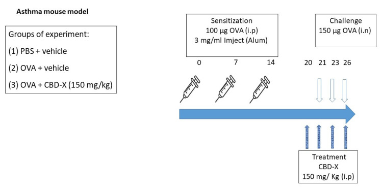
Methods: We evaluated the effects of CBD-X on cells involved in asthma pathogenesis using primary human Th2 cells, neutrophils, and asthma mouse model.

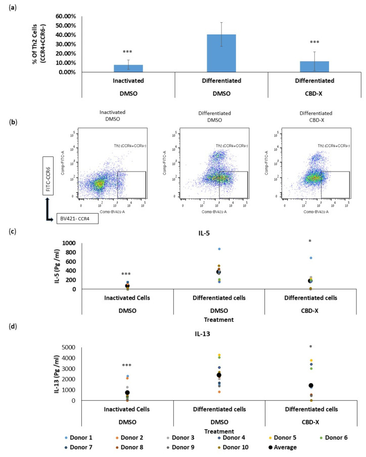
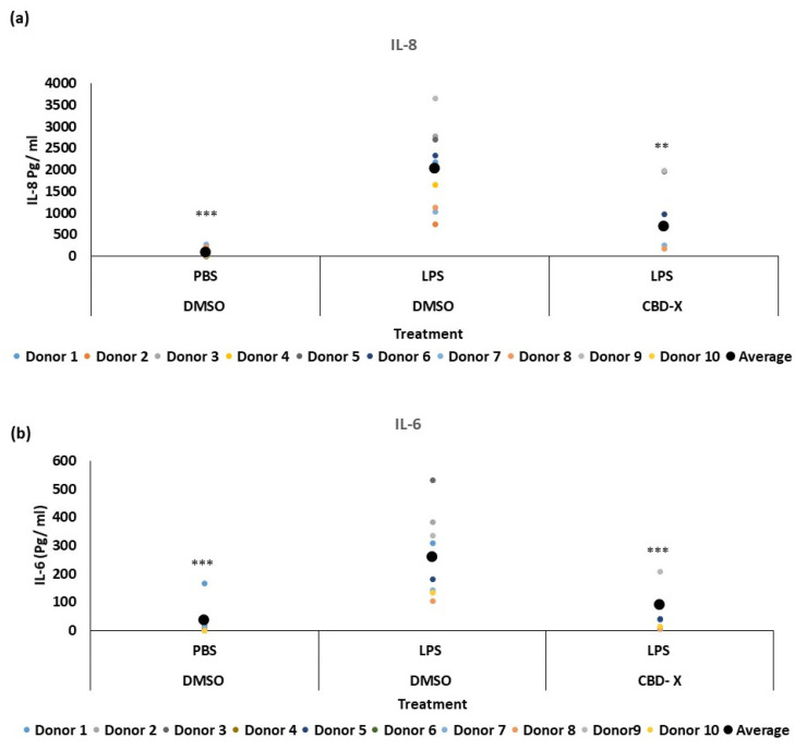
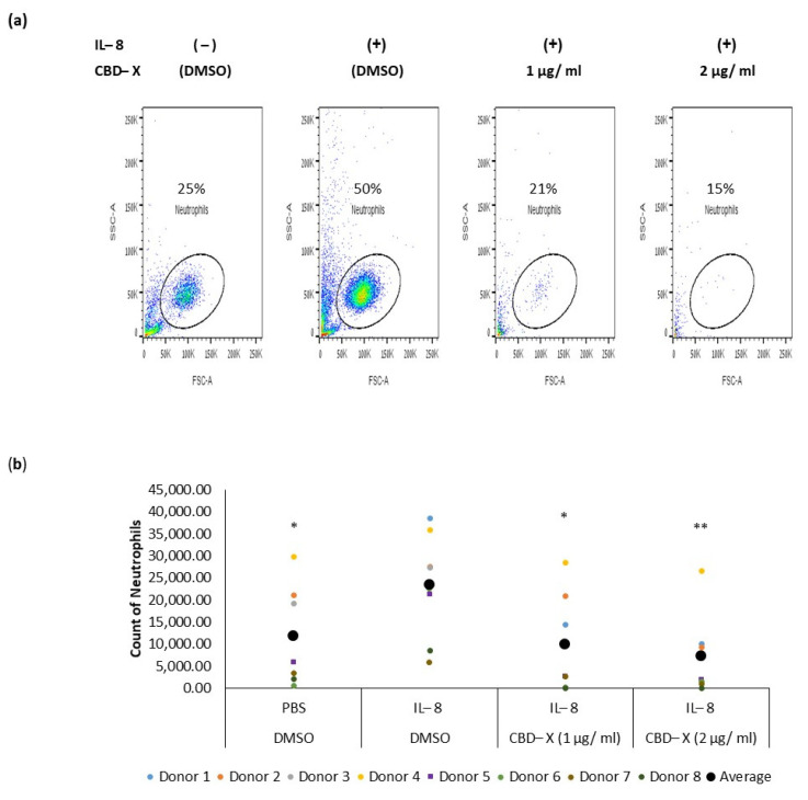
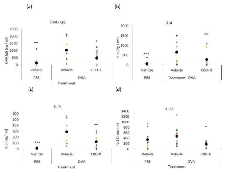
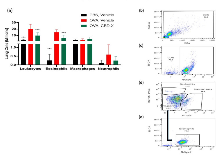
Results: Our findings indicate that CBD-X extract inhibits Th2 differentiation and reduces the secretion of IL-5 and IL-13, which are crucial cytokines in asthma. Additionally, CBD-X significantly reduces pro-inflammatory cytokines IL-8 and IL-6 in neutrophils and impairs their migration, a critical step in airway inflammation. In a murine asthma model, CBD-X administration led to marked downregulation of IgE and pro-asthmatic cytokines, along with reduced leukocyte, eosinophil, and neutrophil infiltration in lung tissues.

Conclusions: These results suggest that CBD-X extract could offer a novel and complementary approach to managing both type 2-high and type 2-low asthma by targeting key inflammatory pathways and modulating immune cell behavior.

Keywords: CBD extracts; IgE; OVA-induced asthmatic mouse model; asthma; cytokines; eosinophil; migration; neutrophil; type 2 helper T cells (Th2).


Conflict of interest statement

This study received funding from Raphael Pharmaceutical Inc. The funders were not involved in any aspect of the study. The authors declare no conflicts of interest.

Figures

Figure 1
Figure 1
CBD-X extract attenuates the differentiation and cytokine release of human T helper 2 (Th2) cells. CBD-X extract was added to isolated differentiated CD4 T cells. Then, Th2 cells were subjected to flow cytometry analysis. CBD-X-treated cells and their control cells were stained with human APC-CD4, BV421- CCR4 and FITC-CCR6. The means and standard deviations of the percentage of Th2 (CCR4+CCR6-) from CD4 population were calculated. (a) The means were calculated from three different experiments of ten healthy donors. Data were analyzed in comparison to differentiated DMSO treatment representative results for CBD-X-treated cells and their control cells are shown (b). Moreover, supernatants were collected and levels of pro-inflammatory cytokines IL5 (c) and IL-13 (d) were detected via ELISA. The means were calculated from healthy donors (black big dot); each dot represents one case. Data were normalized to differentiated cell groups and analyzed via one-way ANOVA (Fisher’s LSD test with values p < 0.05 considered statistically significant, (* p < 0.05, *** p < 0.001).
Figure 2
Figure 2
CBD-X downregulates the secretion of pro-inflammatory cytokines from human neutrophils. Isolated neutrophils were treated with 2 µg/mL CBD-X. Treated cells were activated via 100 ng/mL LPS overnight. Levels of IL-8 and IL-6 (a,b) were detected via ELISA. Each colored dot represents one donor. The means were calculated from healthy donors (black big dot) and each dot represents one case. Data were analyzed via one-way ANOVA (Fisher’s LSD test with values p < 0.05 considered statistically significant, (** p < 0.01, *** p < 0.001).
Figure 3
Figure 3
CBD-X extract inhibits the migration of neutrophils induced by IL-8. CBD-X-treated neutrophils were seeded onto the 3 µm pore polyester membrane in the upper chamber of a 24-well Boyden chamber. Human IL-8 in a concentration of 1 ng/mL was placed in the lower chamber. After three hours, cells that had migrated through the pores into the lower chamber were collected and subjected to flow cytometry analysis. (a) Representative results for migrated neutrophils toward IL-8 after treatment with (1 and 2 µg/mL) CBD-X. The colors in the dot plots represent the quantity of the cells. The lightest color (i.e., blue) would represent the low number of cells. In contrast, green, yellow and red would represent increasing values, with red indicating the highest values. (b) The number of migrated neutrophils. The means were calculated from healthy donors (black big dot) and each dot represents one case. Data were analyzed in comparison to the “IL-8, DMSO” group and analyzed via One-way ANOVA (Fisher’s LSD test with values p < 0.05 considered statistically significant, (* p < 0.05, ** p < 0.01).
Figure 4
Figure 4
CBD-X extract attenuates serum OVA- IgE and BALF cytokine levels in an asthma mouse model. BALB/c mice were induced with OVA and were treated with 150 mg/kg CBD-X extract. Mice were euthanized, blood and lung fluids were collected. Levels of serum OVA- IgE (a) were detected via ELISA. Alternatively, levels of the pro-inflammatory IL-4 (b), IL-5 (c), and IL-13 (d) were also detected in the lung fluids via ELISA. Each colored dot represents one mouse. Standard deviations were calculated as the means of three biologically independent experiments (black big dot); n = 10. Data were analyzed via one-way ANOVA (Fisher’s LSD test with values p < 0.05 considered statistically significant, (* p <0.05, ** p < 0.01, *** p < 0.001).
Figure 5
Figure 5
CBD-X extract inhibits migration of leukocytes to OVA-induced asthma lungs. Mice were induced with OVA-induced asthma and were treated with 150 mg/kg of CBD-X extract. Then, mice were sacrificed and lung fluids were collected. (a) Cells in the lung fluids were stained with anti-mouse APC-CD45, anti-mouse FITC- F480, anti-mouse BV786-LY6G, and PE- Siglec F for flow cytometry analysis. Numbers of leukocytes, eosinophils, macrophages, and neutrophils were calculated in the three different treatments; PBS- vehicle (Black bars), OVA- vehicle (Red bars) and OVA- CBD-X (green bars) (a). The gating strategy is (b) FSC vs. SSC, (c) APC-CD45 vs. SSC, and (d) BV786-LY6G vs. FITC-F4/80. (e) PE- Siglec F vs. SSC out of the negative population gate in (d). The colors in the dot plots represent the quantity of the cells. The lightest color (i.e., blue) would represent the low number of cells. In contrast, green, yellow and red would represent increasing values, with red indicating the highest values. Averages and standard deviations were calculated as the means of two biologically independent experiments, each experiment included five mice per group. Data were analyzed via mixed-effects model (Fisher’s LSD test with values p < 0.05 considered statistically significant, (*** p < 0.001).
Figure 6
Figure 6
CBD-X treatment in a mouse model of OVA-induced asthma. For sensitization, BALB/c mice received an i.p injection of OVA (100 µg) with 3 mg/mL Inject (Alum) (aluminum hydroxide (40 mg/mL) and magnesium hydroxide (40 mg/mL) on days 1, 7, and 14. PBS was injected as a control. The mice were challenged with an (intranasal) i.n injection of OVA (150 µg) or PBS on days 21, 23, and 26. Mice were injected intraperitoneally with 150 mg/kg CBD-X extract or vehicle as a control on days 20, 21, 23, and 26. After 3 h of the last challenge, mice were euthanized and lung fluids were collected using PBS through lavage.

Similar articles

Cited by

References

    1. Song P., Adeloye D., Salim H., Dos Santos J.P., Campbell H., Sheikh A., Rudan I. Global, Regional, and National Prevalence of Asthma in 2019: A Systematic Analysis and Modelling Study. J. Glob. Health. 2022;12:04052. doi: 10.7189/jogh.12.04052. - DOI - PMC - PubMed
    1. Evans C.M., Kim K., Tuvim M.J., Dickey B.F. Mucus Hypersecretion in Asthma: Causes and Effects. Curr. Opin. Pulm. Med. 2009;15:4–11. doi: 10.1097/MCP.0b013e32831da8d3. - DOI - PMC - PubMed
    1. Fehrenbach H., Wagner C., Wegmann M. Airway Remodeling in Asthma: What Really Matters. Cell Tissue Res. 2017;367:551–569. doi: 10.1007/s00441-016-2566-8. - DOI - PMC - PubMed
    1. Chapman D.G., Irvin C.G. Mechanisms of Airway Hyper-responsiveness in Asthma: The Past, Present and yet to Come. Clin. Exp. Allergy. 2015;45:706–719. doi: 10.1111/cea.12506. - DOI - PMC - PubMed
    1. Wu W., Bang S., Bleecker E.R., Castro M., Denlinger L., Erzurum S.C., Fahy J.V., Fitzpatrick A.M., Gaston B.M., Hastie A.T., et al. Multiview Cluster Analysis Identifies Variable Corticosteroid Response Phenotypes in Severe Asthma. Am. J. Respir. Crit. Care Med. 2019;199:1358–1367. doi: 10.1164/rccm.201808-1543OC. - DOI - PMC - PubMed

LinkOut - more resources