. 2024 Oct 20;17(10):1400.
doi: 10.3390/ph17101400.

Hydrogel Containing Propolis: Physical Characterization and Evaluation of Biological Activities for Potential Use in the Treatment of Skin Lesions

Affiliations

Hydrogel Containing Propolis: Physical Characterization and Evaluation of Biological Activities for Potential Use in the Treatment of Skin Lesions

Lindalva Maria de Meneses Costa Ferreira et al. Pharmaceuticals (Basel). .

Abstract

Background: Skin injury affects the integrity of the skin structure and induces the wound healing process, which is defined by a well-coordinated series of cellular and molecular reactions that aim to recover or replace the injured tissue. Hydrogels are a group of promising biomaterials that are able to incorporate active ingredients for use as dressings. This study aimed to synthesize hydrogels with and without propolis extract and evaluate their physical characteristics and biological activities in vitro for potential use as active dressings in the treatment of skin lesions.

Methods: The antifungal [Candida albicans (C. albicans) and Candida tropicalis (C. tropicalis)] and antibacterial [Staphylococcus aureus (S. aureus), Pseudomonas aeruginosas (P. aeruginosas) and Escherichia coli (E. coli)] activity was assessed by the microdilution method in plates and antioxidant potential by the reduction of the phosphomolybdate complex.

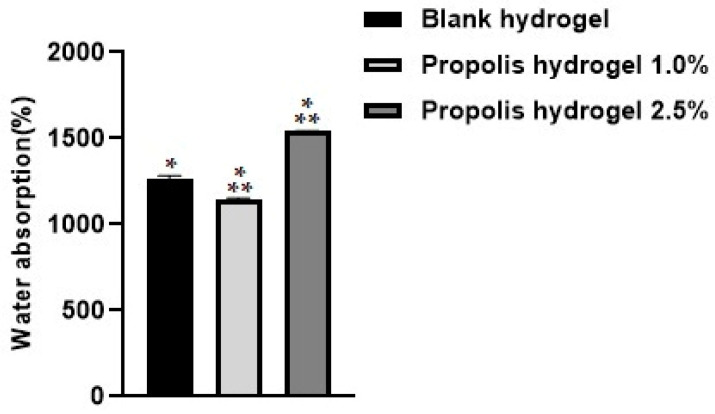
Results: The hydrogels showed good water absorption capacity, high solubility, and high gel fraction, as well as good porosity, water retention, and vapor transmission rates. They revealed a totally amorphous structure. The extract and the hydrogels containing the propolis extract (1.0% and 2.5%) did not inhibit fungal growth. However, they showed antibacterial activity against strains of S. aureus and P. aeruginosas. Regarding the E. coli strain, only the extract inhibited its growth. It showed good antioxidant activity by the evaluation method used.

Conclusions: Therefore, the hydrogels containing propolis extract can be a promising alternative with antibacterial and antioxidant action for use as dressings for the treatment of skin lesions.

Keywords: antimicrobial; antioxidant; biomaterial; chronic wounds; natural product.


Conflict of interest statement

The authors declare no conflicts of interest.

Figures

Figure 1
Figure 1
Water absorption analysis of the blank hydrogel and hydrogels containing propolis extract (1.0% and 2.5%) after 24 h. Results were obtained using ANOVA followed by Tukey’s test. * Significant difference between hydrogels containing propolis extract 1.0% and 2.5% and the blank (p < 0.01). ** Significant difference between the hydrogel containing propolis extract 1.0% and that containing propolis extract 2.5% (p < 0.01).
Figure 2
Figure 2
Water-solubility test performed on blank hydrogel and propolis extract hydrogels (1% and 2.5%). Results were obtained using ANOVA followed by Tukey’s test. NS: non-significant difference between the hydrogel containing propolis extract 1.0% and 2.5% and the blank and non-significant difference between the hydrogel containing propolis extract 1.0% cand that containing propolis extract 2.5%.
Figure 3
Figure 3
Evaluation of the porosity of the blank hydrogel and propolis extract hydrogels (1% and 2.5%). Results were obtained using ANOVA followed by Tukey’s test. NS: non-significant difference between the hydrogel containing propolis extract 1.0% and the blank (p > 0.01). ** Significant difference between the hydrogel containing propolis extract 2.5% and the blank (p < 0.01). *** Significant difference between the hydrogel containing propolis extract 2.5% and that containing propolis extract 1.0% (p < 0.01).
Figure 4
Figure 4
Evaluation of the gel fraction of the blank hydrogel and propolis extract hydrogels (1% and 2.5%). Results were obtained using ANOVA followed by Tukey’s test. NS: non-significant difference between the hydrogels containing propolis extract 1.0% and 2.5% and the blank and non-significant difference between the hydrogel containing propolis extract 1.0% and that containing propolis extract 2.5%.
Figure 5
Figure 5
Evaluation of the water retention capacity of the blank hydrogel and propolis extract hydrogels (1.0% and 2.5%).
Figure 6
Figure 6
Analysis of water loss through the rate of water vapor transmission of the blank hydrogel and propolis extract hydrogels (1.0% and 2.5%).
Figure 7
Figure 7
XRD diffractograms of the blank hydrogel and propolis extract hydrogels. (A) Blank hydrogel, (B) hydrogel containing 1.0% extract, (C) hydrogel containing 2.5% extract. Conditions: Scanning angle 5° ≤ 2θ ≤ 50°, step size 0.02°, and acquisition time of 1 s per step.
Figure 8
Figure 8
Percentage of inhibition in the evaluation of antifungal activity in strains of Candida albicans and Candida tropicalis at different concentrations. (A) Propolis extract, (B) blank hydrogel, (C) hydrogel containing 1.0% extract, (D) hydrogel containing 2.5% extract, and (E) drug used as standard (fluconazole).
Figure 9
Figure 9
Optical density of Candida albicans strains. (A) Propolis extract, (B) white hydrogel, (C) hydrogel containing 1.0% extract, and (D) hydrogel containing 2.5% extract. G.C: growth control (+). S.C: sterility control (−). Results were obtained using ANOVA followed by Tukey’s test. * p < 0.01 and ** p < 0.05.
Figure 10
Figure 10
Optical density of Candida tropicalis strains. (A) Propolis extract, (B) white hydrogel, (C) hydrogel containing 1.0% extract, and (D) hydrogel containing 2.5% extract. G.C: growth control (+). S.C: sterility control (−). Results were obtained using ANOVA followed by Tukey’s test. * p < 0.01 and ** p < 0.05.
Figure 11
Figure 11
Percentage of inhibition in the evaluation of the antibacterial activity against strains of Staphylococcus aureus, Pseudomonas aeruginosa, and Escherichia coli after 24 h of treatment. (A) Propolis extract, (B) white hydrogel, (C) hydrogel containing 1.0% extract, (D) hydrogel containing 2.5% extract, and (E) drug used as standard (penicillin + streptomycin).
Figure 12
Figure 12
Optical density of Staphylococcus aureus strains. (A) Propolis extract, (B) blank hydrogel, (C) hydrogel containing 1.0% extract, and (D) hydrogel containing 2.5% extract. G.C: growth control (+). S.C: sterility control (−). Results were obtained using ANOVA followed by Tukey’s test. * p < 0.01 and ** p < 0.05.
Figure 13
Figure 13
Optical density of Pseudomonas aeroginosa strains from propolis extract, blank hydrogel, and propolis hydrogels. (A) Propolis extract, (B) white hydrogel, (C) hydrogel containing 1.0% extract, and (D) hydrogel containing 2.5% extract. G.C: growth control (+). S.C: sterility control (−). Results were obtained using ANOVA followed by Tukey’s test. * p < 0.01 and ** p < 0.05.
Figure 14
Figure 14
Optical density of Escherichia coli strains from propolis extract, blank hydrogel, and propolis hydrogels. (A) Propolis extract, (B) white hydrogel, (C) hydrogel containing 1.0% extract, and (D) hydrogel containing 2.5% extract. G.C: growth control (+). S.C: sterility control (−). Results were obtained using ANOVA followed by Tukey’s test. * p < 0.01 and ** p < 0.05.
Figure 15
Figure 15
Data obtained in the evaluation of antioxidant activity by reducing the phosphomolybdenum complex in the extract, blank hydrogel, and propolis extract hydrogels (1.0% and 2.5%). AAR a.a: Percentage of antioxidant activity relative to ascorbic acid. Results were obtained using ANOVA followed by Tukey’s test. NS: non-significant difference between propolis extract and the blank, hydrogel containing propolis extract 1.0% and 2.5% and propolis, and non-significant difference between the hydrogel containing propolis extract 1.0% and 2.5% and the blank.

Similar articles

Cited by

References

    1. Permyakova E.S., Solovieva A.O., Sitnikova N., Kiryukhantsev-Korneev P.V., Kutzhanov M.K., Sheveyko A.N., Ignatov S.G., Slukin P.V., Shtansky D.V., Manakhov A.M. Polycaprolactone nanofibers functionalized by fibronectin/gentamicin and implanted silver for enhanced antibacterial properties, cell adhesion, and proliferation. Polymers. 2024;16:261. doi: 10.3390/polym16020261. - DOI - PMC - PubMed
    1. Zhan X., Wang D., Wang H., Chen H., Wu X., Li T., Qi J., Chen T., Wu D., Gao Y. Revitalizing skin repair: Unveiling the healing power of livisin, a natural peptide calcium mimetic. Toxins. 2024;16:21. doi: 10.3390/toxins16010021. - DOI - PMC - PubMed
    1. Zheremyan E.A., Ustiugova A.S., Karamushka N.M., Uvarova A.N., Stasevich E.M., Bogolyubova A.V., Kuprash D.V., Korneev K.V. Breg-mediated immunoregulation in the skin. Int. J. Mol. Sci. 2024;25:583. doi: 10.3390/ijms25010583. - DOI - PMC - PubMed
    1. Julovi S.M., McKelvey K., Minhas N., Chan Y.K.A., Xue M., Jackson C.J. Involvement of PAR-2 in the induction of cell-specific matrix metalloproteinase-2 by activated protein C in cutaneous wound healing. Int. J. Mol. Sci. 2024;25:370. doi: 10.3390/ijms25010370. - DOI - PMC - PubMed
    1. Quiñones-Vico M.I., Fernández-González A., Ubago-Rodríguez A., Moll K., Norrby-Teglund A., Svensson M., Gutiérrez-Fernández J., Torres J.M., Arias-Santiago S. Antibiotics against Pseudomonas aeruginosa on human skin cell lines: Determination of the highest non-cytotoxic concentrations with antibiofilm capacity for wound healing strategies. Pharmaceutics. 2024;16:117. doi: 10.3390/pharmaceutics16010117. - DOI - PMC - PubMed

LinkOut - more resources