

Nanoscale Extracellular Vesicle-Enabled Liquid Biopsy: Advances and Challenges for Lung Cancer Detection
- PMID: 39459055
- PMCID: PMC11509190
- DOI: 10.3390/mi15101181
Nanoscale Extracellular Vesicle-Enabled Liquid Biopsy: Advances and Challenges for Lung Cancer Detection
Abstract
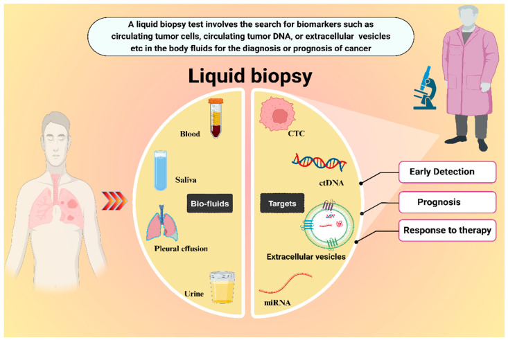
Lung cancer is responsible for the death of over a million people worldwide every year. With its high mortality rate and exponentially growing number of new cases, lung cancer is a major threat to public health. The high mortality and poor survival rates of lung cancer patients can be attributed to its stealth progression and late diagnosis. For a long time, intrusive tissue biopsy has been considered the gold standard for lung cancer diagnosis and subtyping; however, the intrinsic limitations of tissue biopsy cannot be overlooked. In addition to being invasive and costly, it also suffers from limitations in sensitivity and specificity, is not suitable for repeated sampling, provides restricted information about the tumor and its molecular landscape, and is inaccessible in several cases. To cope with this, advancements in diagnostic technologies, such as liquid biopsy, have shown great prospects. Liquid biopsy is an innovative non-invasive approach in which cancer-related components called biomarkers are detected in body fluids, such as blood, urine, saliva and others. It offers a less invasive alternative with the potential for applications such as routine screening, predicting treatment outcomes, evaluating treatment effectiveness, detecting residual disease, or disease recurrence. A large number of research articles have indicated extracellular vesicles (EVs) as ideal biomarkers for liquid biopsy. EVs are a heterogeneous collection of membranous nanoparticles with diverse sizes, contents, and surface markers. EVs play a critical role in pathophysiological states and have gained prominence as diagnostic and prognostic biomarkers for multiple diseases, including lung cancer. In this review, we provide a detailed overview of the potential of EV-based liquid biopsy for lung cancer. Moreover, it highlights the strengths and weaknesses of various contemporary techniques for EV isolation and analysis in addition to the challenges that need to be addressed to ensure the widespread clinical application of EV-based liquid biopsies for lung cancer. In summary, EV-based liquid biopsies present interesting opportunities for the development of novel diagnostic and prognostic platforms for lung cancer, one of the most abundant cancers responsible for millions of cancer-related deaths worldwide.
Keywords: biomarkers; circulating tumor DNA; circulating tumor cells; exosomes; extracellular vesicles; liquid biopsy; lung cancer; non-invasive diagnosis.
Conflict of interest statement
The authors declare that they have no conflicts of interest.
Figures


References
-
- Mehta A., Barreto G. Non-invasive approaches for lung cancer diagnosis. Indian J. Thorac. Cardiovasc. Surg. 2018;34:11–19. doi: 10.1007/s12055-017-0600-4. - DOI
Publication types
LinkOut - more resources
Full Text Sources
