Review
. 2024 Sep 26;14(10):1234.
doi: 10.3390/life14101234.

The Microbiota-Gut-Brain Axis and Neurological Disorders: A Comprehensive Review

Affiliations
Review

The Microbiota-Gut-Brain Axis and Neurological Disorders: A Comprehensive Review

Mohammed M Nakhal et al. Life (Basel). .

Abstract

Microbes have inhabited the earth for hundreds of millions of years longer than humans. The microbiota-gut-brain axis (MGBA) represents a bidirectional communication pathway. These communications occur between the central nervous system (CNS), the enteric nervous system (ENS), and the emotional and cognitive centres of the brain. The field of research on the gut-brain axis has grown significantly during the past two decades. Signalling occurs between the gut microbiota and the brain through the neural, endocrine, immune, and humoral pathways. A substantial body of evidence indicates that the MGBA plays a pivotal role in various neurological diseases. These include Alzheimer's disease (AD), autism spectrum disorder (ASD), Rett syndrome, attention deficit hyperactivity disorder (ADHD), non-Alzheimer's neurodegeneration and dementias, fronto-temporal lobe dementia (FTLD), Wilson-Konovalov disease (WD), multisystem atrophy (MSA), Huntington's chorea (HC), Parkinson's disease (PD), multiple sclerosis (MS), amyotrophic lateral sclerosis (ALS), temporal lobe epilepsy (TLE), depression, and schizophrenia (SCZ). Furthermore, the bidirectional correlation between therapeutics and the gut-brain axis will be discussed. Conversely, the mood of delivery, exercise, psychotropic agents, stress, and neurologic drugs can influence the MGBA. By understanding the MGBA, it may be possible to facilitate research into microbial-based interventions and therapeutic strategies for neurological diseases.

Keywords: microbiota; microbiota–gut–brain axis; neurological disorders and neurodegenerative diseases; psychotropic agents.


Conflict of interest statement

The authors declare that the research was conducted in the absence of any commercial or financial relationships that could be construed as a potential conflict of interest.

Figures

Figure 1
Figure 1
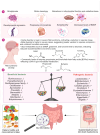
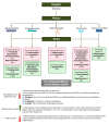
Brain disorder classifications. Neurological disorders are divided in this review into developmental disabilities and metabolic disorders, neurodegenerative disorders (NDDs), immune-mediated nervous system diseases, non-communicable neurological disorders, and mental (behavioural) disorders.
Figure 2
Figure 2
Altered gut microbiota in Rett syndrome. Increased levels of harmful bacteria such as Clostridium and a reduction in beneficial bacteria like Bifidobacterium and Lactobacillus reflect an imbalance in the gut microbial community in patients with Rett syndrome (RTT), indicating an association with gastrointestinal (GI) symptoms such as constipation, bloating, and abdominal pain. Immune modulation, metabolic changes, and the production of neuroactive compounds are mechanisms that may influence the gut–brain axis in RTT patients.
Figure 3
Figure 3
The link between gut microbiota and their metabolites with lipid dysregulation in Alzheimer disease (AD). The involvement of short-chain fatty acids (SCFAs), phospholipids, and other metabolites generated by gut bacteria in the synthesis and degradation of structural and functional lipids in cells might play a role in the progression and deterioration of AD pathology.
Figure 4
Figure 4
Therapeutic and non-pharmacological interventions employed to enhance cognitive functions in Alzheimer disease (AD). Microbiome-based therapies for AD include probiotics, prebiotics, synbiotics, postbiotics, and faecal material transplantation (FMT). The pathogenic proteins amyloid beta and tau contribute to hypothalamic–pituitary–adrenal (HPA) axis dysregulation, which results in synaptotoxicity and amyloidosis. These interventions aim to modulate the gut microbiota, improve AD symptoms, and modulate plaque-associated microglial functions. However, the beneficial and side effects of these approaches on the central nervous system (CNS), the endocrine system, and the immune system have yet to be fully studied.
Figure 5
Figure 5
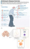
Symptomatology and pathological hallmarks of Parkinson disease (PD). (1) Clinical features and symptoms of PD; (2) biomarkers and neuroimaging evidence that identifies alpha-synuclein deposits.
Figure 6
Figure 6
Pathogenesis of synaptic dysfunction in multiple sclerosis (MS). Healthy neurons in MS display normal morphology and function, with intact myelin sheaths that facilitate rapid and efficient signal transmission. In contrast, damaged neurons often exhibit demyelination, where the protective myelin coating is disrupted or lost, leading to impaired signal conduction.
Figure 7
Figure 7
Altered serotonin transporter levels in patients with MS. Tryptophan (TRP) is converted into serotonin (5-HT) through a series of chemical reactions, with the enzyme tryptophan hydroxylase (TPH) playing a key role. Some of the most studied sites of 5-HT action include the gastrointestinal (GI) system, cerebral cortex, and hypothalamus. Activation of the 5-HT1A receptor in clusters of differentiation 4 (CD4+) cells increases interleukin-10 (IL-10) production [164]. Conversely, activation of the 5-HT3 receptor stimulates T cells to produce inflammatory mediators like interleukin-6 (IL-6) and interleukin-7 (IL-17). This demonstrates how serotonin synthesis and its varied roles are intricately connected to the gut microbiota, especially in the context of sickness behaviour.
Figure 8
Figure 8
Microorganisms’ alteration in multiple sclerosis (MS). This diagram highlights specific changes in the microbiome associated with the disease and their impacts on the immune system, both beneficial and detrimental.
Figure 9
Figure 9
The central nervous system (CNS) and peripheral nervous system (PNS) bidirectionally communicate with the gut microbiota. (A) The peripheral nervous system includes the cranial and spinal nerves, in particular, the ganglia nerves that extend beyond the CNS, and the autonomic nervous system, which consists of the sympathetic and parasympathetic branches as demonstrated in. The CNS plasticity includes many cellular and anatomical mechanisms, reflecting synaptic efficacy and synaptic redundancy. The creation of new neurons in the CNS is known as neurogenesis, while synaptogenesis involves the formation of synapses that facilitate neuronal communication. The autonomic nervous system which communicates with internal organs and glands has a flexibility that reflects the integrity of central and peripheral systems, incorporating the adaptation support to environmental demands and thereby serving as a key indicator of neuroplasticity. The digestive tract possesses its distinct nervous system called the ENS. Neurons found in certain nerve clusters transmit sensory information from the body’s outer regions to the CNS. (B) The ENS comprises plexuses that consist of neurons. Individual enteric neurons function either as intrinsic afferent, efferent, motor neurons, or interneurons. The myenteric plexus resides between the longitudinal and circular muscle layers. The small intestine alone houses approximately 100 million neurons, making the ENS the largest collection of neurons and glia outside the brain.
Figure 10
Figure 10
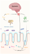
Correlation between the brain and gut microbiome in major depressive disorder (MDD). Stressful circumstances can disrupt the delicate balance of the gut microbiota, resulting elevated levels of proinflammatory cytokines, particularly interleukin-6 (IL-6) and interferon gamma (IFN-γ), and reduced levels of short-chain fatty acids (SCFAs), and weaken the integrity of the gut, facilitating the migration of bacteria (leaky gut). An imbalance in the kynurenine pathway results from increased levels of inflammatory cytokines stimulate the action of indoleamine 2, 3-dioxygenase (IDO), which interferes with the synthesis of protective metabolites such as kynurenic acid (KYNA). As a result, compromising the blood–brain barrier (BBB) increases inflammation in brain tissue and causes astrocyte atrophy and microglial activation. Probiotics and prebiotics have been shown to modify the gut microbiota and improve intestinal barrier function, which in turn indirectly reduces BBB permeability, toxic metabolites from the kynurenine pathway, and inflammatory cytokines. LBS: gut-derived lipopolysaccharides.
Figure 11
Figure 11
Dopamine agonists and adjunctive pharmacotherapies for Parkinson’s disease (PD). This illustration elucidates how dopamine agonists are potentially adjunctive treatments later in the disease course, along with other approved pharmacologic options for alleviating motor symptoms associated with the disease through the microbiome, contributing to a comprehensive management strategy for the disease. Abbreviations: Levodopa (L-Dopa), catechol-O-methyl-transferase (COMT), Monoamine oxidase-B (MAO-B). Green arrows indicate an increase and red arrows indicate a decrease [254,256,259].
Figure 12
Figure 12
Psychotropic drugs, microbiome composition and mental disorders. Psychotropic drugs work by influencing the levels and activity of neurotransmitters, the chemicals in the brain that transmit signals between nerve cells. The goal of these medications is to correct imbalances in neurotransmitter levels, thereby alleviating symptoms and improving the quality of life for individuals with mental health conditions. This figure explains how psychotropic agents are connected to the gut microbiome, altering its bioavailability. Abbreviations: selective serotonin reuptake inhibitors (SSRIs), gamma-aminobutyric acid (GABA) [228].
Figure 13
Figure 13
The impact of delivery mode on infant gut microbiota. The mode of delivery significantly alters neonatal gut microbiota. C-sections can affect intestinal epithelial cell activation and immune system development. Conversely, vaginal birth exposes infants to beneficial maternal microbiota, promoting a balanced immune system, gut function, and short-chain fatty acids (SCFAs).
Figure 14
Figure 14
The effect of exercise on the microbiome. (A) Individuals who engage in at least three hours of exercise per week exhibited elevated levels of butyrate producing bacteria, including Akkermansia muciniphila, Faecalibacterium prausnitzii, and Roseburia hominis. (B) The comparison between moderate and high intensity exercise. The abundance of Bifidobacterium and butyrate-producing bacteria, including Lachnospira eligens and Enterococcus spp., was greater in individuals who participated in lower intensity exercises. Additionally, obese and overweight males who practiced high intensity exercise presented with reduction in faecal and serum levels of branched-chain amino acids and aromatic amino acids. They also had increased faecal propionate, gamma-aminobutyric acid (GABA), and short-chain fatty acids (SCFAs). Abbreviation: branched-chain amino acids (BCAAs).

References

    1. Rhee S.H., Pothoulakis C., Mayer E.A. Principles and Clinical Implications of the Brain-Gut-Enteric Microbiota Axis. Nat. Rev. Gastroenterol. Hepatol. 2009;6:306–314. doi: 10.1038/nrgastro.2009.35. - DOI - PMC - PubMed
    1. Cryan J.F., O’Mahony S.M. The Microbiome-Gut-Brain Axis: From Bowel to Behavior. Neurogastroenterol. Motil. 2011;23:187–192. doi: 10.1111/j.1365-2982.2010.01664.x. - DOI - PubMed
    1. De Palma G., Collins S.M., Bercik P. The Microbiota-Gut-Brain Axis in Functional Gastrointestinal Disorders. Gut Microbes. 2014;5:419–429. doi: 10.4161/gmic.29417. - DOI - PMC - PubMed
    1. Sudo N., Chida Y., Aiba Y., Sonoda J., Oyama N., Yu X., Kubo C., Koga Y. Postnatal Microbial Colonization Programs the Hypothalamic–Pituitary–Adrenal System for Stress Response in Mice. J. Physiol. 2004;558:263–275. doi: 10.1113/jphysiol.2004.063388. - DOI - PMC - PubMed
    1. Gareau M.G., Wine E., Rodrigues D.M., Cho J.H., Whary M.T., Philpott D.J., MacQueen G., Sherman P.M. Bacterial Infection Causes Stress-Induced Memory Dysfunction in Mice. Gut. 2011;60:307–317. doi: 10.1136/gut.2009.202515. - DOI - PubMed

LinkOut - more resources