

Between Life and Death: Sea Urchin Embryos Undergo Peculiar DNA Fragmentation after Exposure to Vanadium, Cadmium, Gadolinium, and Selenium
- PMID: 39459596
- PMCID: PMC11508963
- DOI: 10.3390/life14101296
Between Life and Death: Sea Urchin Embryos Undergo Peculiar DNA Fragmentation after Exposure to Vanadium, Cadmium, Gadolinium, and Selenium
Abstract
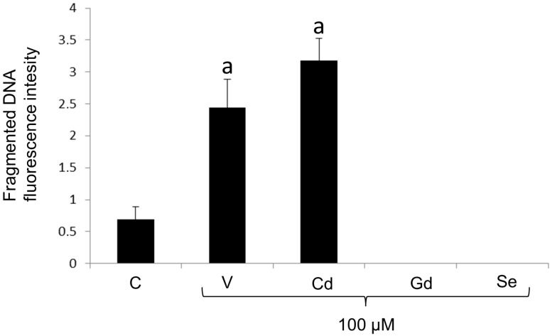
Exogenous DNA damage represents one of the most harmful outcomes produced by environmental, physical, or chemical agents. Here, a comparative analysis of DNA fragmentation was carried out on Paracentrotus lividus sea urchin embryos exposed to four common pollutants of the marine environment: vanadium, cadmium, gadolinium and selenium. Using the terminal deoxynucleotidyl transferase dUTP nick end labeling (TUNEL) assay, fragmented DNA was quantified and localized in apoptotic cells mapping whole-mount embryos. This is the first study reporting how different chemicals are able to activate distinctive apoptotic features in sea urchin embryos, categorized as follows: (i) cell-selective apoptosis, showing DNA fragmentation restricted to a subset of extremely damaged cells, acting as an embryo survival mechanism; or (ii) total apoptosis, with fragmented DNA widespread throughout the cells of the entire embryo, leading to its death. Also, this is the first report of the effects of Se exposure on P. lividus sea urchin embryos. These data confirm the TUNEL assay as the most suitable test to study DNA fragmentation in the sea urchin embryo model system. Taken together, this research highlights embryos' ability to find alternative pathways and set physiological limits for development under stress conditions.
Keywords: DNA fragmentation; TUNEL assay; apoptosis; marine ecotoxicology; metal pollution; sea urchin embryo.
Conflict of interest statement
The authors declare no conflicts of interest.
Figures
Similar articles
-
Cadmium induces an apoptotic response in sea urchin embryos.Cell Stress Chaperones. 2007 Spring;12(1):44-50. doi: 10.1379/csc-229r.1. Cell Stress Chaperones. 2007. PMID: 17441506 Free PMC article.
-
Toxicity of Vanadium during Development of Sea Urchin Embryos: Bioaccumulation, Calcium Depletion, ERK Modulation and Cell-Selective Apoptosis.Int J Mol Sci. 2022 Jun 2;23(11):6239. doi: 10.3390/ijms23116239. Int J Mol Sci. 2022. PMID: 35682917 Free PMC article.
-
Autophagy as a defense strategy against stress: focus on Paracentrotus lividus sea urchin embryos exposed to cadmium.Cell Stress Chaperones. 2016 Jan;21(1):19-27. doi: 10.1007/s12192-015-0639-3. Epub 2015 Sep 11. Cell Stress Chaperones. 2016. PMID: 26362931 Free PMC article. Review.
-
Environmentally relevant cadmium concentrations affect development and induce apoptosis of Paracentrotus lividus larvae cultured in vitro.Cell Biol Toxicol. 2008 Dec;24(6):603-10. doi: 10.1007/s10565-008-9066-x. Epub 2008 Mar 6. Cell Biol Toxicol. 2008. PMID: 18322810
-
Cadmium stress effects indicating marine pollution in different species of sea urchin employed as environmental bioindicators.Cell Stress Chaperones. 2019 Jul;24(4):675-687. doi: 10.1007/s12192-019-01010-1. Epub 2019 Jun 5. Cell Stress Chaperones. 2019. PMID: 31165437 Free PMC article. Review.
References
-
- Naselli F., Cardinale P.S., Volpes S., Martino C., Cruciata I., Valenti R., Luparello C., Caradonna F., Chiarelli R. An alternative approach of TUNEL assay to specifically characterize DNA fragmentation in cell model systems. Histochem. Cell Biol. 2024;162:429–442. doi: 10.1007/s00418-024-02306-9. - DOI - PMC - PubMed
Grants and funding
LinkOut - more resources
Full Text Sources
