Review
. 2024 Sep 27;16(10):1528.
doi: 10.3390/v16101528.

The Assembly of HTLV-1-How Does It Differ from HIV-1?

Affiliations
Review

The Assembly of HTLV-1-How Does It Differ from HIV-1?

Dominik Herrmann et al. Viruses. .

Abstract

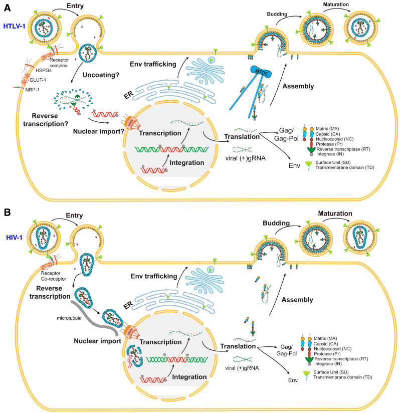
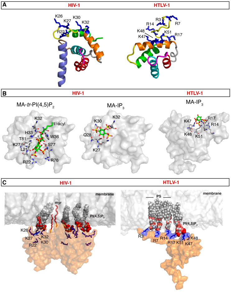
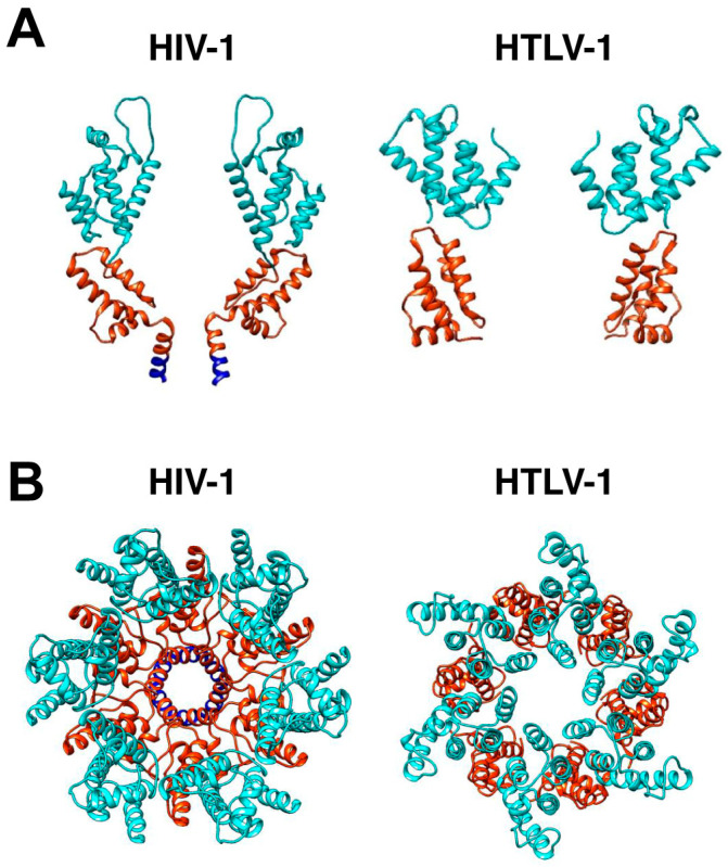
Retroviral assembly is a highly coordinated step in the replication cycle. The process is initiated when the newly synthesized Gag and Gag-Pol polyproteins are directed to the inner leaflet of the plasma membrane (PM), where they facilitate the budding and release of immature viral particles. Extensive research over the years has provided crucial insights into the molecular determinants of this assembly step. It is established that Gag targeting and binding to the PM is mediated by interactions of the matrix (MA) domain and acidic phospholipids such as phosphatidylinositol 4,5-bisphosphate (PI(4,5)P2). This binding event, along with binding to viral RNA, initiates oligomerization of Gag on the PM, a process mediated by the capsid (CA) domain. Much of the previous studies have focused on human immunodeficiency virus type 1 (HIV-1). Although the general steps of retroviral replication are consistent across different retroviruses, comparative studies revealed notable differences in the structure and function of viral components. In this review, we present recent findings on the assembly mechanisms of Human T-cell leukemia virus type 1 and highlight key differences from HIV-1, focusing particularly on the molecular determinants of Gag-PM interactions and CA assembly.

Keywords: Gag polyprotein; capsid (CA); human T-cell leukemia virus type 1 (HTLV-1); human immunodeficiency virus type 1 (HIV-1); matrix (MA); phosphatidylinositol 4,5-bisphosphate (PI(4,5)P2); plasma membrane (PM).


Conflict of interest statement

The authors declare no conflicts of interest.

Figures

Figure 1
Figure 1
HTLV-1 and HTLV-1 replication cycles. (A) Mature HTLV-1 virion attaches to the host cell receptor complex containing NRP-1, GLUT-1, and HSPGs. The mechanisms of reverse transcription and uncoating have long been thought to occur in the cytoplasm but recent advances on the mechanisms of HIV-1 reverse transcription and uncoating (below) raised similar questions about other retroviruses, including HTLV-1. Subsequent nuclear import and integration into the host genome yields the provirus. Transcription and translation produce Gag, Gag-Pol, Env, accessory proteins, and viral gRNA. Gag is trafficked to the PM for assembly via the MTOC, while Env is post-translationally processed and trafficked to the cell surface through the ER and Golgi apparatus. Virus assembly and maturation yield a new, infectious virus. (B) Mature HIV-1 virion attaches to the host CD4 receptor and co-receptors (CCR5 or CXCR4). The virus core is then transported to the nucleus via microtubules, a process that appears to be accompanied by reverse transcription. Recent studies indicated that CA core uncoating occurs in the nucleus near the integration sites. Transcription and translation produce Gag, Gag-Pol, Env, accessory proteins, and viral gRNA. Gag is then trafficked to the PM for assembly, while Env is post-translationally processed and trafficked to the cell surface through the ER and Golgi apparatus. Virus assembly and maturation yield a new, infectious virus.
Figure 2
Figure 2
HTLV-1 genome and RNA transcripts. Genome encodes for Gag, Pro, Pol, Env, Tax, Rex, and pX genes. pX region contains genes of Rex, Tax, p30, p12, p13, and HBZ (antisense transcript). mRNA transcripts are 5′-capped and 3′-polyadenylated. Alternative splicing yields mRNA for Env, Tax, Rex, p12, p13, p30, and HBZ.
Figure 3
Figure 3
MA-membrane binding models for HIV-1 and HTLV-1. (A) Structures of HIV-1 myrMA (PDB code 2H3I) and HTLV-1 myr(–)MA (PDB code 7M1W). Structures highlight the HBR implicated in membrane binding (blue sticks). For HIV-1 myrMA, the following residues are not shown for clarity: myr group, residues 2–3 and 115–132. For HTLV-1 myr(–)MA, the following residues are not shown for clarity: 1–2 and 94–99. (B) Surface representation of the HIV-1 myrMA structure (PDB code 2H3I) highlighting residues that exhibited substantial chemical shift changes upon binding of tr-P(4,5)P2 (PDB code 2H3V) and IP3 (left and middle, respectively). Structures are viewed in identical orientations. The structure of HTLV-1 myr(–)MA bound to IP3 is shown on the right. (C) Models of HIV-1 myrMA and HTLV-1 myr(–)MA bound to membrane showing interactions between PI(4,5)P2 and/or PS and the HBR. Membrane bilayer was constructed by CHARMM-GUI [169].
Figure 4
Figure 4
HIV-1 and HTLV-1 Gag hexamer structures. (A) The two HIV-1 CA molecules are displayed on the side of the hexamer, with CANTD in cyan and CACTD in orange. HIV-1 SP1 domains are shown in blue. The PDB codes are HIV-1 (5L93) [109], HTLV-1 CANTD (8PUG) [183], and HTLV-1 CACTD (8PUH) [183]. The cross-section of the HTLV-1 Gag lattice reconstruction map suggests a distinctive arrangement of the CANTD and CACTD compared to HIV-1. (B) Shown is the top view of the HIV-1 hexamer structure, which was generated by fitting HIV-1 CA (5L93) into the EM density of the immature HIV-1 lattice (EMD: 4017). The top view of the HTLV-1 Gag hexamer structure shown was generated by fitting CANTD and CACTD separately into the EM density of the immature HTLV-1 CA lattice (EMD: 17942). The flexible linker between HTLV-1 CANTD and CACTD is unstructured and is therefore not shown.
Figure 5
Figure 5
Comparison of Env CT. (A) Schematic representation of the gp41 subunits, indicating the lengths of their respective cytoplasmic tails (25 and 150 amino acids for HTLV-1 and HIV-1, respectively. (B) Secondary structure representation of the HIV-1 gp41CT protein based on the NMR data [192]. (C) HIV-1 Env incorporation is mediated by interaction between the MA domain of the Gag lattice and gp41CT. For HTLV-1, the CT appears to contain functional motifs that play important roles in cell-to-cell infection and syncytium formation.
Figure 6
Figure 6
Comparison of MA lattices based on structural data. (A) Schematic representation of the myrMA lattice in the immature and mature states based on the cryo-ET data [170]. The trimer–trimer interactions are mediated by the N-terminal domain in the vicinity of the myr group, while the PI(4,5)P2 binding pocket is empty. In the mature myrMA lattice, PI(4,5)P2 is bound to the cleft and myrMA trimer–trimer interactions are formed by the HBR and PI(4,5)P2. (B) Schematic illustration of the myrMA lattice based on the X-ray structure of myrMA. In this lattice, myrMA–myrMA interaction at the trimer–trimer interface is mediated by the N-terminal residues. Of note, myrMA–myrMA interaction at the trimer–trimer interface places the myr groups (red) in juxtaposition. The HBR and PI(4,5)P2 binding cleft are also shown. Hexagons and triangles denote C6 and C3 symmetry, respectively.

Similar articles

References

    1. Ruscetti F.W., Morgan D.A., Gallo R.C. Functional and morphologic characterization of human T cells continuously grown in vitro. J. Immunol. 1977;119:131–138. doi: 10.4049/jimmunol.119.1.131. - DOI - PubMed
    1. Morgan D.A., Ruscetti F.W., Gallo R. Selective in vitro growth of T lymphocytes from normal human bone marrows. Science. 1976;193:1007–1008. doi: 10.1126/science.181845. - DOI - PubMed
    1. Bobrow S.N., Smith R.G., Reitz M.S., Gallo R.C. Stimulated normal human lymphocytes contain a ribonuclease-sensitive DNA polymerase distinct from viral RNA-directed DNA polymerase. Proc. Natl. Acad. Sci. USA. 1972;69:3228–3232. doi: 10.1073/pnas.69.11.3228. - DOI - PMC - PubMed
    1. Smith R.G., Abrell J.W., Lewis B.J., Gallo R.C. Serological analysis of human deoxyribonucleic acid polymerases. Preparation and properties of antiserum to deoxyribonucleic acid polymerase I from human lymphoid cells. J. Biol. Chem. 1975;250:1702–1709. doi: 10.1016/S0021-9258(19)41750-X. - DOI - PubMed
    1. Poiesz B.J., Ruscetti F.W., Gazdar A.F., Bunn P.A., Minna J.D., Gallo R.C. Detection and isolation of type-C retrovirus particles from fresh and cultured lymphocytes of a patient with cutaneous T-cell lymphoma. Proc. Natl. Acad. Sci. USA. 1980;75:7415–7419. doi: 10.1073/pnas.77.12.7415. - DOI - PMC - PubMed

Publication types

Substances

LinkOut - more resources