. 2024 Oct 11;24(20):6549.
doi: 10.3390/s24206549.

A Microbial Cocaine Bioreporter

Affiliations

A Microbial Cocaine Bioreporter

Anne-Kathrin Grimm et al. Sensors (Basel). .

Abstract

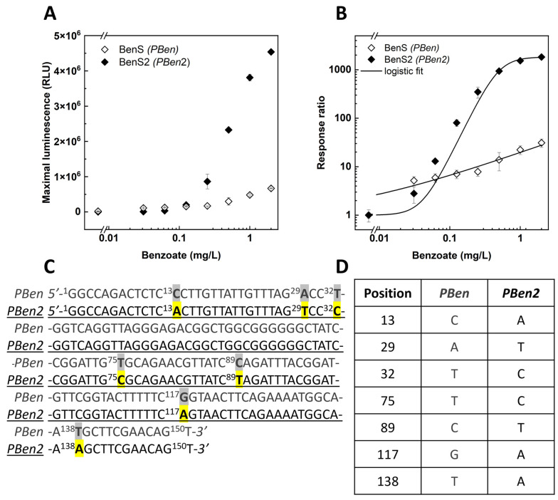
The continuous emergence of new illegal compounds, particularly psychoactive chemicals, poses significant challenges for current drug detection methods. Developing new protocols and kits for each new drug requires substantial time, effort, and dedicated manpower. Whole-cell bacterial bioreporters have been proven capable of detecting diverse hazardous compounds in both laboratory and field settings, identifying not only single compounds but also chemical families. We present the development of a microbial bioreporter for the detection of cocaine, the nervous system stimulant that is the second-most widely used illegal drug in the US. Escherichia coli was transformed with a plasmid containing a bacterial luxCDABEG bioluminescence gene cassette, activated by a cocaine-responsive signaling cascade. The engineered bioreporter is demonstrated to be a sensitive and specific first-generation detection system for cocaine, with detection thresholds of 17 ± 8 μg/L and 130 ± 50 μg/L in a buffer solution and in urine, respectively. Further improvement of the sensor's performance was achieved by altering the nucleotide sequence of the PBen gene promoter, the construct's sensing element, using accelerated site-directed evolution. The applicability of ready-to-use paper strips with immobilized bioreporter cells was demonstrated for cocaine detection in aqueous solutions.

Keywords: Escherichia coli; Pseudomonas putida; ben operon; bioluminescence; cocaine; cocaine esterase; microbial bioreporters.


Conflict of interest statement

The authors declare no conflicts of interest.

Figures

Figure 1
Figure 1
(A) Schemes of the two plasmids constituting the cocaine detection circuit. Plasmid I harbors the cocaine esterase gene (cocE) and the transcription factor benR. Plasmid II harbors the P. leiognathi luxCDABEG gene cassette controlled by the BenR-inducible promotor PBen. (B) Cocaine detection signaling cascade. Following cocaine cleavage by cocaine esterase (CocE) to ecgonine methyl ester (EcME) and benzoate (1), the latter complexes with the transcription factor BenR (2) and activates the PBen promotor (3). This drives the expression of the luxCDABEG genes, leading to a dose-dependent luminescence signal (4). (C) Schematic illustration of the benzoate sensor plasmid (plasmid III), which harbors the transcription factor gene benR along with the PBAD promoter and its regulator gene araC. The luxCDABEG gene cassette is under the control of PBen. (D) In the presence of arabinose, benR is expressed (1); the BenR protein forms a complex with a benzoate molecule (2), initiating transcription of luxCDABEG by activation of the PBen promotor (3), leading to quantifiable luminescence (4).
Figure 2
Figure 2
Luminescent cocaine detection by CocS cells in liquid medium. (A) Time course of luminescence development following exposure of CocS cells to the indicated cocaine concentrations. (B) Dose dependency of the luminescent signal (maximal values over a 16 h exposure), displayed both as signal intensity (◊) and as the response ratio (♦). Luminescence intensity values (mean ± SEM, n = 6) in both panels are presented in the plate reader’s arbitrary relative luminescence units (RLUs). LOD was determined using the response ratio plus three times the standard deviation of the background. (C) Luminescence of CocS cells 5.5 h post exposure to different cocaine concentrations. Image was captured by a Sony Alpha 7sII camera (4 s exposure, f-number 2, ISO 40,000). (D) Increase in pixel intensity of images shown in C compared with the cocaine-free control. Intensities were determined using the mean gray value measurement in ImageJ. Data shown represent mean ± SD (area = 3268 pixels) with incorporation of error propagation. LOD was set to be three standard deviations above the background. (E) Response specificity: luminescence development over time during exposure of CocS cells in the presence of different compounds (mean ± SEM, n = 2)—5 mg/L cocaine, 5 mg/L benzoate, 40 mg/L fentanyl, 40 mg/L ketamine, 0.1 mg/L THC, and 2 mg/L CBD. (F) Functionality in human urine: maximal luminescence presented both as signal intensity (◊) and as the response ratio (♦) of two individual samples from different donors (mean ± SEM, n = 2) with cocaine concentrations ranging from 0.152 to 5 mg/L.
Figure 3
Figure 3
Induction of lyophilized CocS cells on paper strips by cocaine (30 min exposure, 30 °C). (A) Luminescence, imaged 5 h after induction using a Sony Alpha 7sII camera (15 s exposure, f-number = 2, ISO 40,000). (B) Increase in pixel intensity (compared with the cocaine-free control) of the images in panel A, analyzed using ImageJ software (version 1.54i, March 2024) which provided both the mean gray value and standard deviation within the bacterial spot region. The figure presents data from two independent experiments after storage at 4 °C. Values are presented in mean ± SEM.
Figure 4
Figure 4
Induction of BenS sensor strain by benzoate. Bacteria were grown overnight at 37 °C and 200 rpm and diluted ×1/150 2 h prior to benzoate induction in the presence (A,B) or absence (C) of 6.6 mM arabinose. Luminescence (mean ± SEM, n = 3) was measured every 10 min at 30 °C. Luminescence values are in the plate reader’s arbitrary relative light units (RLUs). Panels (B,D) present the maximal luminescence (RLU) and calculated response ratio as a function of benzoate concentration in the presence or absence of arabinose, respectively.
Figure 5
Figure 5
Responses to benzoate of the BenS2 sensor, harboring the mutated version of the PBen promoter (PBen2). (A) Maximal luminescence response (mean ± SEM, n = 3) of BenS2 and BenS to benzoate. (B) Response ratios of the two sensors. (C) Gene sequence of the PBen and PBen2 promoters, with the seven point mutations highlighted. (D) Mutations in PBen2 and their positions.
Figure 6
Figure 6
Cocaine detection (as response ratio) by CocS and CocS2 sensor strains.

References

    1. van der Meer J.R., Belkin S. Where microbiology meets microengineering: Design and applications of reporter bacteria. Nat. Rev. Microbiol. 2010;8:511–522. doi: 10.1038/nrmicro2392. - DOI - PubMed
    1. Yagi K. Applications of whole-cell bacterial sensors in biotechnology and environmental science. Appl. Microbiol. Biotechnol. 2007;3:1251–1258. doi: 10.1007/s00253-006-0718-6. - DOI - PubMed
    1. Elad T., Almog R., Yagur-Kroll S., Levkov K., Melamed S., Shacham-Diamand Y., Belkin S. Online monitoring of water toxicity by use of bioluminescent reporter bacterial biochips. Environ. Sci. Technol. 2011;45:8536–8544. doi: 10.1021/es202465c. - DOI - PubMed
    1. Reifferscheid G., Buchinger S. Cell-based genotoxicity testing: Genetically modified and genetically engineered bacteria in environmental genotoxicology. Adv. Biochem. Eng. Biotechnol. 2010;118:85–111. - PubMed
    1. Belkin S., Yagur-Kroll S., Kabessa Y., Korouma V., Septon T., Anati Y., Zohar-Perez C., Rabinovitz Z., Nussinovitch A., Agranat A.J. Remote detection of buried landmines using a bacterial sensor. Nat. Biotechnol. 2017;35:308–310. doi: 10.1038/nbt.3791. - DOI - PubMed

LinkOut - more resources