. 2024 Dec;65(12):3717-3731.
doi: 10.1111/epi.18166. Epub 2024 Oct 26.

Mouse models of Slc35a2 brain mosaicism reveal mechanisms of mild malformations of cortical development with oligodendroglial hyperplasia in epilepsy

Affiliations

Mouse models of Slc35a2 brain mosaicism reveal mechanisms of mild malformations of cortical development with oligodendroglial hyperplasia in epilepsy

Hyojung Yoon et al. Epilepsia. 2024 Dec.

Abstract

Objective: Brain somatic variants in SLC35A2 were recently identified as a genetic marker for mild malformations of cortical development with oligodendroglial hyperplasia in epilepsy (MOGHE). The role of SLC35A2 in cortical development and the contributions of abnormal neurons and oligodendrocytes to seizure activity in MOGHE remain largely unexplored.

Methods: Here, we generated a novel Slc35a2 floxed allele, which we used to develop two Slc35a2 conditional knockout mouse lines targeting (1) the Emx1 dorsal telencephalic lineage (excitatory neurons and glia) and (2) the Olig2 lineage (oligodendrocytes). We examined brain structure, behavior, and seizure activity.

Results: Knockout of Slc35a2 from the Emx1 lineage, which targets both cortical neurons and oligodendrocytes, resulted in early lethality and caused abnormal cortical development, increased oligodendroglial cell density, early onset seizures, and developmental delays akin to what is observed in patients with MOGHE. By tracing neuronal development with 5-Ethynyl-2'-deoxyuridine (EdU) birthdating experiments, we found that Slc35a2 deficiency disrupts corticogenesis by delaying radial migration of neurons from the subventricular zone. To discern the contributions of oligodendrocytes to these phenotypes, we knocked out Slc35a2 from the Olig2 lineage. This recapitulated the increased oligodendroglial cell density and resulted in abnormal electroencephalographic activity, but without a clear seizure phenotype, suggesting Slc35a2 deficiency in neurons is required for epileptogenesis.

Significance: This study presents two novel Slc35a2 conditional knockout mouse models and characterizes the effects on brain development, behavior, and epileptogenesis. Together, these results demonstrate a direct causal role for SLC35A2 in MOGHE-like phenotypes, including a critical role in neuronal migration during brain development, and identify neurons as key contributors to SLC35A2-related epileptogenesis.

Keywords: brain mosaicism; glycosylation; malformation of cortical development; pediatrics.


Conflict of interest statement

T.A.B. is cofounder of BehaviorCloud. The remaining authors have no conflicts of interest. The authors confirm that we have read the Journal's position on issues involved in ethical publication and affirm that this report is consistent with those guidelines.

Figures

FIGURE 1
FIGURE 1
Emx1‐Cre‐mediated Slc35a2 conditional knockout (cKO) mice have developmental delays and impaired growth and survival. (A) Emx1‐Cre mice were crossed with Slc35a2 floxed mice to produce forebrain‐specific loss of SLC35A2 from excitatory neurons and glia (blue). Cx, cortex; Hp, hippocampus; OB, olfactory bulb. (B) Slc35a2 gene expression measured by real‐time quantitative polymerase chain reaction was significantly reduced in the cortices of cKO mice compared to floxed controls (n = 4 per group; one‐way analysis of variance [ANOVA], F 3,12 = 21.16, p < .0001, Bonferroni post hoc comparisons shown). (C) SLC35A2 protein expression measured by Western blot was significantly reduced in the cortices of cKO male mice compared to floxed male controls (n = 3–4 per group; one‐way ANOVA, F 3,11 = 4.228, p < .05, Bonferroni post hoc comparisons shown). (D, E) Male cKO mice were smaller (D) and had significantly reduced body weight (E) beginning at postnatal day 9 (n = 7–24 mice per group; mixed‐effect model [genotype × time] with Tukey post hoc comparisons, p < .05). (F) Both male and female cKO mice had reduced survival compared to floxed controls (p < .0001), and hemizygous males had reduced survival compared to heterozygous females (p < .0001; n = 32–88 per group; Kaplan–Meier survival differences evaluated by the log‐rank test). (G–J) Neonatal cKO mice exhibited developmental delays including delayed eye opening (G), delayed ear twitch flattening reflex (H), delayed auditory startle reflex (I), and abnormal hind limb clasping reflex (J; n = 6–23 per group; two‐tailed t‐test). *p < .05, **p < .01, ****p < .0001. Data are shown as mean ± SEM.
FIGURE 2
FIGURE 2
Cortical architecture and oligodendroglial cell density in Emx1‐Cre‐mediated Slc35a2 conditional knockout (cKO) mice. (A) Representative images of coronal sections through the postnatal day 8 brain stained with 4,6‐diamidino‐2‐phenylindole (DAPI). Lines indicate measurements of cortex (ctx) thickness and corpus callosum (cc) thickness. (B) Both female and male cKO mice had reduced cortex thickness (one‐way analysis of variance [ANOVA] with Bonferroni post hoc comparisons, F 3,16 = 31.51, p < .0001; post hoc p < .001 in females and p < .0001 in males). (C) No differences in corpus callosum thickness were observed. (D–F) Representative images of coronal sections through the cortex stained with layer markers BRN2 (layers II/III), SATB2 (layers II–IV), and CTIP2 (layers V/VI). Cortex was divided into six equal bins from the ventricle to cortical surface to analyze distribution of positive immunofluorescent area. (G) BRN2 staining was more widely distributed in female and male cKO mice compared to floxed controls, with significantly decreased BRN2 positivity in bin 1 of both sexes and increased BRN2 staining found in bin 2 (n = 3–4 per group, 2–3 sections per brain; two‐way ANOVA with Bonferroni post hoc comparisons; interaction of genotype × cortical layer, F 15,96 = 12.55, p < .0001; post hoc comparisons shown). (H) SATB2 staining was reduced in male cKO mice compared to floxed controls in bin 2 and bin 3 (n = 3–4 per group, 2–3 sections per brain; two‐way ANOVA with Bonferroni post hoc comparisons; effect of genotype, F 3,90 = 3.618, p < .05; post hoc comparisons shown). (I) No significant differences were observed in CTIP2 staining across cortical bins. (J) OLIG2 immunoreactivity was quantified in an region of interest containing the cortex–white matter junction (blue box). (K) OLIG2‐positive cell density was significantly increased in the cortex–white matter junction of male and female cKO mice compared to floxed controls (n = 3–4 per group, 2–3 sections per brain; two‐tailed t‐tests, p < .05). Data are shown as mean ± SEM. *p < .05, ***p < .001, ****p < .0001.
FIGURE 3
FIGURE 3
Neuronal migration is delayed in Emx1‐Cre‐mediated Slc35a2 conditional knockout (cKO) mice. (A) To determine whether a defect in neuronal migration was responsible for the changes in cortical layering observed within the cortex, newborn neurons destined for layer II/III were labeled by EdU injection at embryonic day 15.5 (E15.5) and brains were subsequently examined at postnatal day 1 (P1) or P8. (B, C) EdU signal was more prevalent in the white matter (WM) and ventricular zone (VZ) compared to cortex in cKO mice at P1, whereas most signal was found in the outer cortex in floxed control mice (n = 3 per group, 2–3 sections per brain; two‐way analysis of variance [ANOVA] with Bonferroni post hoc tests; interaction of genotype × cortical layer, F 6,24 = 64.99, p < .0001; post hoc comparisons shown). (D, E) By P8, EdU signal was concentrated in the outer cortex in both floxed control and cKO mice, indicating a delay in neuronal migration (n = 3 mice per group, 2–4 sections per brain; two‐way ANOVA with Bonferroni post hoc tests, no significance). Data are shown as mean ± SEM. *p < .05, ***p < .001, ****p < .0001. DAPI, 4,6‐diamidino‐2‐phenylindole.
FIGURE 4
FIGURE 4
Emx1‐Cre‐mediated Slc35a2 conditional knockout (cKO) mice exhibit spontaneous seizure activity. (A) Representative electroencephalographic (EEG) traces over 5 min of recording from floxed control and Emx1‐Cre‐mediated Slc35a2 cKO mice between 15 and 25 weeks of age. cKO mice exhibited spontaneous bursts of electrographic activity correlating with behavioral abnormalities (highlighted by black bars) and abnormal interictal spike discharges. (B) Highlighted regions of EEG activity are shown in more detail. Emx1‐Cre‐mediated Slc35a2 cKO mice demonstrated rhythmic ictal spiking with clear onset and offset. (C) An average of 26 electrographically confirmed seizures were observed in cKO mice during the 72‐h video‐EEG recordings compared to none in the floxed controls (Mann–Whitney test, *p < .05). (D) These events coincided with behavioral signs of seizure, including leg/head jerking (score = 2–3) and clonic activity (e.g., falling to the side; score = 4–5; Mann–Whitney test, **p < .01). Male and female mice are pooled. Data shown are as mean ± SEM.
FIGURE 5
FIGURE 5
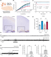
Olig2‐Cre‐mediated Slc35a2 conditional knockout (cKO) mice reveal contributions of oligodendrocytes to abnormal electroencephalographic (EEG) activity. (A) Olig2‐Cre mice were crossed with Slc35a2 floxed mice to delete SLC35A2 from oligodendrocytes. (B) Olig2‐Cre‐mediated Slc35a2 cKO mice had reduced body mass compared to floxed controls (n = 7–15 mice per group; one‐way analysis of variance with Tukey multiple comparisons; effect of genotype, F 3,814 = 170.2, p < .0001, post hoc p < .05 in +/y vs. −/y males day 7–21, +/+ vs. −/+ females day 11–21, −/+ females vs. −/y males day 13–15 and day 19–21). (C) Olig2‐Cre‐mediated Slc35a2 cKO males, but not females, had significantly reduced survival. Kaplan–Meier survival differences were evaluated by the log‐rank test (p < .01). (D) OLIG2 immunoreactivity was quantified in the cortex–white matter junction (blue box). (E) OLIG2‐positive cell density was significantly increased in the cortex–white matter junction of male cKO mice (n = 3–5 per group, 2–4 sections per brain; two‐tailed t‐tests, p < .05). (F) Representative EEG traces over 5 min of recording from floxed control and Olig2‐Cre‐mediated Slc35a2 cKO mice between 15 and 25 weeks of age. The same floxed control mice were used for both Figure 4 and Figure 5 comparisons, replotted here for direct comparison. cKO mice exhibit spontaneous bursts of >10 s. Ictal spiking activity correlating with behavioral abnormalities (highlighted by black bars) and abnormal spike discharges are shown. (G) Highlighted regions of EEG activity are shown in more detail. Olig2‐Cre‐mediated Slc35a2 cKOs exhibited bursts of irregular spikes. (H) An average of 13 ictal discharges were observed over the 72‐h recording period in Olig2‐Cre‐mediated Slc35a2 cKO mice (Mann–Whitney test, p < .05). (I) Behavioral correlates of these events corresponded to mild twitching or jerking (score = 2–3; Mann–Whitney test, p < .05). Male and female mice are pooled. Data are shown as mean ± SEM. *p < .05.

References

    1. Najm I, Lal D, Alonso Vanegas M, Cendes F, Lopes‐Cendes I, Palmini A, et al. The ILAE consensus classification of focal cortical dysplasia: An update proposed by an ad hoc task force of the ILAE diagnostic methods commission. Epilepsia. 2022;63:1899–1919. - PMC - PubMed
    1. Winawer MR, Griffin NG, Samanamud J, Baugh EH, Rathakrishnan D, Ramalingam S, et al. Somatic SLC35A2 variants in the brain are associated with intractable neocortical epilepsy. Ann Neurol. 2018;83:1133–1146. - PMC - PubMed
    1. Bonduelle T, Hartlieb T, Baldassari S, Sim NS, Kim SH, Kang HC, et al. Frequent SLC35A2 brain mosaicism in mild malformation of cortical development with oligodendroglial hyperplasia in epilepsy (MOGHE). Acta Neuropathol Commun. 2021;9:3. - PMC - PubMed
    1. Sprong H, Degroote S, Nilsson T, Kawakita M, Ishida N, van der Sluijs P, et al. Association of the Golgi UDP‐galactose transporter with UDP‐galactose:ceramide galactosyltransferase allows UDP‐galactose import in the endoplasmic reticulum. Mol Biol Cell. 2003;14:3482–3493. - PMC - PubMed
    1. Wiktor M, Wiertelak W, Maszczak‐Seneczko D, Balwierz PJ, Szulc B, Olczak M. Identification of novel potential interaction partners of UDP‐galactose (SLC35A2), UDP‐N‐acetylglucosamine (SLC35A3) and an orphan (SLC35A4) nucleotide sugar transporters. J Proteome. 2021;249:104321. - PubMed

Substances